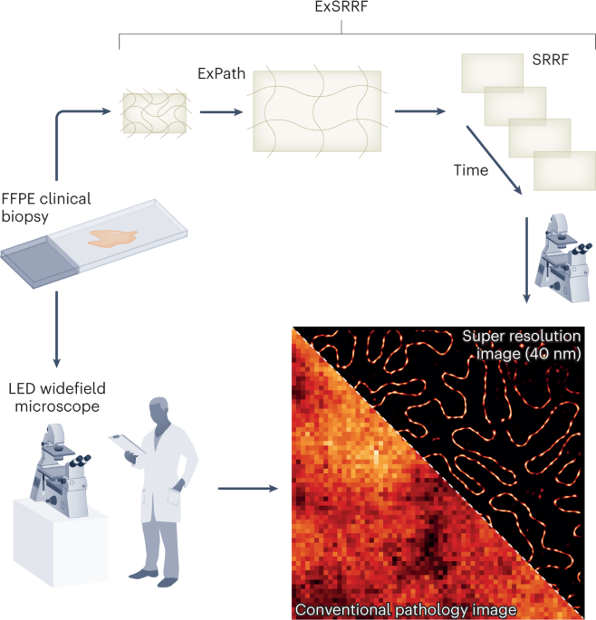
When something goes awry with our body, we often turn to pathology, the study of how diseases develop and progress. It encompasses both the microscopic examination of cellular changes that take place during disease, as well as the molecular mechanisms underlying those changes. Pathologists use a variety of techniques to examine tissues and organs from patients to determine the cause of disease, helping clinicians treat patients more effectively. Pathologists typically use optical microscopes to examine tissues, observing morphological variations that could indicate disease. Most pathologists have access to LED-based widefield microscopes that allow large-scale morphological examination with relatively poor resolution. This can prove less than ideal, as many disease-causing changes occur at the nanoscale, subcellular level. Current optical super-resolution methods, including structured illumination, stimulated emission depletion, and stochastic optical reconstruction microscopy, require hardware that is prohibitively expensive for most pathology laboratories and has a steep learning curve to operate. Now, writing in Nature Nanotechnology, Kylies et al. have reported expansion enhanced super-resolution radial fluctuations (or ExSRRF)1, an addition to the expansion microscopy toolkit that eases these burdens on clinicians and aims to make trivial the super-resolution examination of pathological samples.
- SEO Powered Content & PR Distribution. Get Amplified Today.
- Platoblockchain. Web3 Metaverse Intelligence. Knowledge Amplified. Access Here.
- Source: https://www.nature.com/articles/s41565-023-01318-1
- :is
- 1
- 2022
- a
- access
- addition
- aims
- AL
- Anchor
- and
- AS
- At
- body
- CAN
- Cause
- Changes
- click
- clinicians
- could
- Current
- curve
- Determine
- develop
- Disease
- diseases
- during
- Eases
- effectively
- emission
- encompasses
- enhanced
- Ether (ETH)
- expansion
- expensive
- fluctuations
- For
- from
- Goes
- Hardware
- Have
- helping
- How
- HTTPS
- ideal
- Imaging
- in
- Including
- indicate
- IT
- large-scale
- learning
- Level
- LINK
- low-cost
- make
- many
- methods
- Microscopy
- molecular
- more
- most
- Nature
- of
- on
- operate
- pathology
- patients
- Place
- plato
- Plato Data Intelligence
- PlatoData
- poor
- Progress
- Prove
- relatively
- Reported
- require
- Resolution
- something
- Strategy
- structured
- Study
- Take
- techniques
- that
- The
- These
- tissues
- to
- toolkit
- treat
- TURN
- typically
- underlying
- use
- variety
- WELL
- with
- writing
- zephyrnet