05 جون 2023 (نانورک نیوز) کینسر کے سرجن جلد ہی نئے امیجنگ ایجنٹوں کی بدولت سرجری کے دوران ٹیومر کے بارے میں مزید مکمل نظریہ حاصل کر سکتے ہیں جو ایک ہی وقت میں متعدد بائیو مارکروں کو روشن کر سکتے ہیں، یونیورسٹی آف الینوائے اربانا-شیمپین کے محققین کی رپورٹ۔ سرخ خون کے خلیوں کی جھلیوں میں لپٹے ہوئے فلوروسینٹ نینو پارٹیکلز، موجودہ طبی طور پر منظور شدہ رنگوں سے بہتر ٹیومر کو نشانہ بناتے ہیں اور سرجیکل لائٹ کے صرف ایک شعاع کے جواب میں دو الگ الگ سگنل خارج کر سکتے ہیں، یہ خصوصیت ڈاکٹروں کو ٹیومر کی سرحدوں میں فرق کرنے اور میٹاسٹیٹک کینسر کی شناخت کرنے میں مدد دے سکتی ہے۔ . امیجنگ ایجنٹوں کو بائیو انسپائرڈ کیمروں کے ساتھ ملایا جا سکتا ہے، جسے محققین نے پہلے سرجری کے دوران حقیقی وقت کی تشخیص کے لیے تیار کیا تھا، ریسرچ گروپ کے رہنما وکٹر گریو، الیکٹریکل اور کمپیوٹر انجینئرنگ کے الینوائے کے پروفیسر نے کہا۔ جرنل میں ایک نئی تحقیق میں ACS نانو ("Cell-membrane coated nanoparticles for tumor delineation and qualitative estimation of cancer biomarkers at single wavelength excitation in murine and phantom models")، محققین نے ٹیومر فینٹمز میں اپنے نئے دوہری سگنل والے نینو پارٹیکلز کا مظاہرہ کیا - 3D ماڈل جو ٹیومر اور ان کے گردونواح کی خصوصیات کی نقل کرتے ہیں - اور زندہ چوہوں میں۔
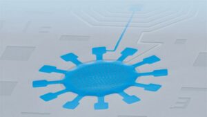
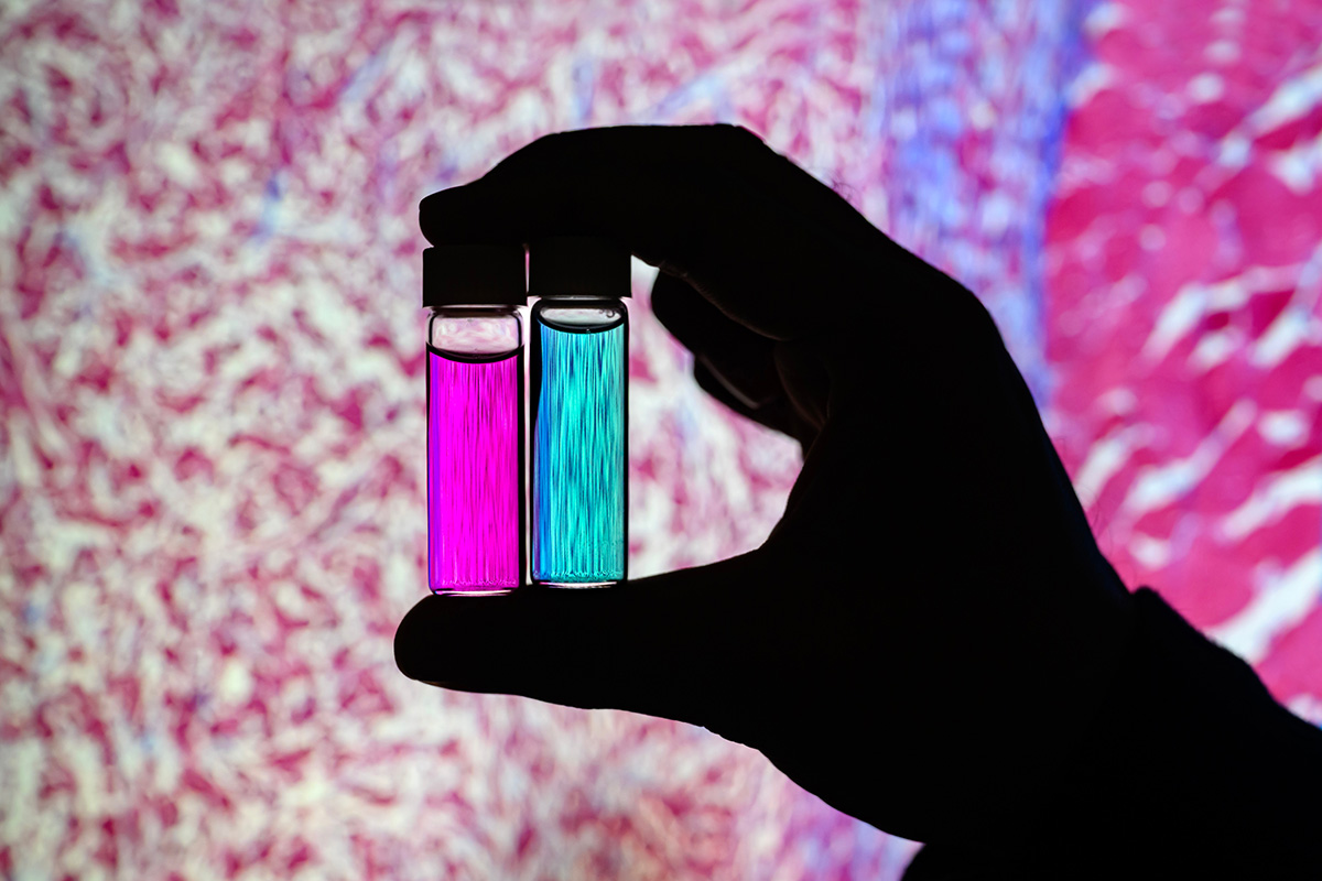 Researcher Indrajit Srivastava holds solutions of nanoparticles that can target two cancer biomarkers, giving off two distinct signals when lit by one fluorescent wavelength. This could give surgeons a more complete picture of a tumor and guide operating-room decisions. In the background is a microscopic scan of a tissue sample. (Image: Fred Zwicky)
“If you want to find all the cancer, imaging one biomarker is not enough. It could miss some tumors. If you introduce a second or a third biomarker, the likelihood of removing all cancer cells increases, and the likelihood of a better outcome for the patients increases.” said Gruev, who also is a professor in the Carle Illinois College of Medicine. “Multiple-targeted drugs and imaging agents are a recent trend, and our group is driving the trend hard because we have the camera technology that can image multiple signals at once.”
Traditionally, a surgeon removes a tumor and sends it to a pathologist for assessment, a process that can take hours to days, said Illinois postdoctoral researcher Indrajit Srivastava, the first author of the paper. As research has moved toward real-time diagnostics, several challenges have prevented wide application: Many tumor-targeted imaging agents only minimally reach their tumor targets, instead being quickly cleared from the blood stream and accumulating in the liver, Srivastava said.
“A few people before us have used nanoparticles coated with red blood cells and found they circulate longer – a few days. We saw the same thing in our mice: The membrane-coated nanoparticles circulated longer in blood, with reduced uptake in the liver. Because they were circulating longer, more of the imaging agents accumulated in the tumors, giving us a stronger fluorescent signal,” Srivastava said.
An artist’s rendering of membrane-wrapped nanoparticles binding to markers on a cancer cell and emitting colored light.
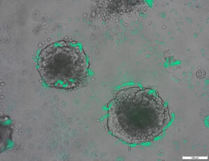
The two biomarkers targeted by the new imaging agents include one that is prevalent in early cancer and one that is prevalent in late-stage cancer, which is more likely to be metastatic. The researchers found that the probes were effective at distinguishing cancerous tissue from healthy tissue, as well as distinguishing the two signals from each other.
“This is appealing for surgical application, as it could help determine where exactly to make the cut. Having multiple signals gives a more overall picture of the tumor. And it could tell a surgeon, ‘This may be metastatic, you may want to be more aggressive in your removal.’” Srivastava said.
Only needing one wavelength of laser light to elicit multiple signals is another benefit for surgical applications, as it makes the instrumentation much more compact than those requiring multiple lasers for each needed wavelength, Gruev said.
The researchers plan to develop more tumor-imaging agents that target multiple markers, and to move forward with further preclinical and clinical studies using their dual-signal dyes with surgical goggles they have developed.
“In this battle for ensuring we remove all the cancer cells during surgery, we need investments both in the imaging camera technology and in the tumor targeting agents,” Gruev said.
Researcher Indrajit Srivastava holds solutions of nanoparticles that can target two cancer biomarkers, giving off two distinct signals when lit by one fluorescent wavelength. This could give surgeons a more complete picture of a tumor and guide operating-room decisions. In the background is a microscopic scan of a tissue sample. (Image: Fred Zwicky)
“If you want to find all the cancer, imaging one biomarker is not enough. It could miss some tumors. If you introduce a second or a third biomarker, the likelihood of removing all cancer cells increases, and the likelihood of a better outcome for the patients increases.” said Gruev, who also is a professor in the Carle Illinois College of Medicine. “Multiple-targeted drugs and imaging agents are a recent trend, and our group is driving the trend hard because we have the camera technology that can image multiple signals at once.”
Traditionally, a surgeon removes a tumor and sends it to a pathologist for assessment, a process that can take hours to days, said Illinois postdoctoral researcher Indrajit Srivastava, the first author of the paper. As research has moved toward real-time diagnostics, several challenges have prevented wide application: Many tumor-targeted imaging agents only minimally reach their tumor targets, instead being quickly cleared from the blood stream and accumulating in the liver, Srivastava said.
“A few people before us have used nanoparticles coated with red blood cells and found they circulate longer – a few days. We saw the same thing in our mice: The membrane-coated nanoparticles circulated longer in blood, with reduced uptake in the liver. Because they were circulating longer, more of the imaging agents accumulated in the tumors, giving us a stronger fluorescent signal,” Srivastava said.
An artist’s rendering of membrane-wrapped nanoparticles binding to markers on a cancer cell and emitting colored light.
The two biomarkers targeted by the new imaging agents include one that is prevalent in early cancer and one that is prevalent in late-stage cancer, which is more likely to be metastatic. The researchers found that the probes were effective at distinguishing cancerous tissue from healthy tissue, as well as distinguishing the two signals from each other.
“This is appealing for surgical application, as it could help determine where exactly to make the cut. Having multiple signals gives a more overall picture of the tumor. And it could tell a surgeon, ‘This may be metastatic, you may want to be more aggressive in your removal.’” Srivastava said.
Only needing one wavelength of laser light to elicit multiple signals is another benefit for surgical applications, as it makes the instrumentation much more compact than those requiring multiple lasers for each needed wavelength, Gruev said.
The researchers plan to develop more tumor-imaging agents that target multiple markers, and to move forward with further preclinical and clinical studies using their dual-signal dyes with surgical goggles they have developed.
“In this battle for ensuring we remove all the cancer cells during surgery, we need investments both in the imaging camera technology and in the tumor targeting agents,” Gruev said.
- SEO سے چلنے والا مواد اور PR کی تقسیم۔ آج ہی بڑھا دیں۔
- پلیٹوآئ اسٹریم۔ ویب 3 ڈیٹا انٹیلی جنس۔ علم میں اضافہ۔ یہاں تک رسائی حاصل کریں۔
- ایڈریین ایشلے کے ساتھ مستقبل کا نقشہ بنانا۔ یہاں تک رسائی حاصل کریں۔
- PREIPO® کے ساتھ PRE-IPO کمپنیوں میں حصص خریدیں اور بیچیں۔ یہاں تک رسائی حاصل کریں۔
- ماخذ: https://www.nanowerk.com/news2/biotech/newsid=63117.php
- : ہے
- : ہے
- : نہیں
- :کہاں
- $UP
- 10
- 12
- 3d
- 7
- 8
- 9
- a
- جمع ہے
- ایجنٹ
- جارحانہ
- تمام
- بھی
- an
- اور
- ایک اور
- اپیل
- درخواست
- ایپلی کیشنز
- نقطہ نظر
- کی منظوری دے دی
- کیا
- AS
- تشخیص
- At
- مصنف
- پس منظر
- جنگ
- BE
- بیم
- کیونکہ
- اس سے پہلے
- کیا جا رہا ہے
- فائدہ
- بہتر
- بائنڈنگ
- بائیومارکر
- خون
- سرحدوں
- دونوں
- by
- کیمرہ
- کیمروں
- کر سکتے ہیں
- کینسر
- کینسر کے خلیات
- خلیات
- سینٹر
- چیلنجوں
- گردش
- کلینکل
- طبی ٹیسٹ
- قریب
- کالج
- مل کر
- مکمل
- کمپیوٹر
- کمپیوٹر انجینئرنگ
- سکتا ہے
- موجودہ
- کٹ
- تاریخ
- دن
- فیصلے
- demonstrated,en
- اس بات کا تعین
- ترقی
- ترقی یافتہ
- مختلف
- ممتاز
- ڈاکٹروں
- ڈرائیونگ
- منشیات
- کے دوران
- ہر ایک
- ابتدائی
- موثر
- انجنیئرنگ
- کافی
- کو یقینی بنانے ہے
- بالکل
- نمایاں کریں
- خصوصیات
- چند
- مل
- پہلا
- کے لئے
- آگے
- ملا
- سے
- مزید
- حاصل کرنے
- دے دو
- فراہم کرتا ہے
- دے
- گروپ
- رہنمائی
- ہاتھ
- ہارڈ
- ہے
- ہونے
- صحت مند
- مدد
- مدد
- انعقاد
- کی ڈگری حاصل کی
- کلی
- HOURS
- HTTPS
- شناخت
- if
- ایلی نوائے
- روشن
- تصویر
- امیجنگ
- in
- شامل
- اضافہ
- کے بجائے
- متعارف کرانے
- سرمایہ کاری
- IT
- جرنل
- فوٹو
- صرف
- صرف ایک
- لیزر
- lasers
- رہنما
- روشنی
- امکان
- رہتے ہیں
- لیور
- اب
- بنا
- بناتا ہے
- بہت سے
- مئی..
- دوا
- چوہوں
- مشرق
- ماڈل
- زیادہ
- منتقل
- آگے بڑھو
- بہت
- ایک سے زیادہ
- ضرورت ہے
- ضرورت
- ضرورت ہے
- نئی
- of
- بند
- on
- ایک بار
- ایک
- صرف
- or
- دیگر
- ہمارے
- نتائج
- مجموعی طور پر
- کاغذ.
- مریضوں
- لوگ
- پریت
- تصویر
- منصوبہ
- پلاٹا
- افلاطون ڈیٹا انٹیلی جنس
- پلیٹو ڈیٹا
- موجودہ
- پہلے
- عمل
- ٹیچر
- قابلیت
- جلدی سے
- تک پہنچنے
- اصل وقت
- احساس
- حال ہی میں
- ریڈ
- کم
- ہٹانے
- ہٹا
- کو ہٹانے کے
- رینڈرنگ
- رپورٹ
- تحقیق
- ریسرچ گروپ
- محقق
- محققین
- جواب
- کہا
- اسی
- اسکین
- دوسری
- بھیجتا ہے
- کئی
- اشارہ
- سگنل
- ایک
- حل
- کچھ
- اسی طرح
- سٹریم
- مضبوط
- مطالعہ
- مطالعہ
- سرجری
- جراحی
- لے لو
- لینے
- ہدف
- ھدف بنائے گئے
- ھدف بندی
- اہداف
- ٹیکنالوجی
- بتا
- سے
- شکریہ
- کہ
- ۔
- ان
- وہ
- بات
- تھرڈ
- اس
- ان
- کرنے کے لئے
- کی طرف
- روایتی طور پر
- رجحان
- ٹرائلز
- دو
- یونیورسٹی
- us
- استعمال کیا جاتا ہے
- کا استعمال کرتے ہوئے
- لنک
- چاہتے ہیں
- we
- اچھا ہے
- تھے
- جب
- جس
- ڈبلیو
- وسیع
- ساتھ
- کام
- لپیٹ
- آپ
- اور
- زیفیرنیٹ
سے زیادہ نانوورک
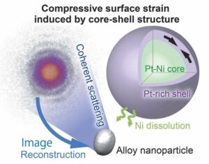
پلاٹینم شیل کے ساتھ نکل پلاٹینم نانوسکل کور آکسیجن کے مالیکیولوں کو مفید آئنوں میں توڑ دیتا ہے۔
ماخذ نوڈ: 2788122
ٹائم اسٹیمپ: جولائی 27، 2023
ڈی این اے کی مرمت کی دریافت بائیو ٹیکنالوجی کو بہتر بنا سکتی ہے (ڈبلیو/ویڈیو)
ماخذ نوڈ: 1987323
ٹائم اسٹیمپ: مارچ 2، 2023
سیل کو نئی حالت میں انجینئر کرنے کے لیے زیادہ موثر تجرباتی ڈیزائن
ماخذ نوڈ: 2914286
ٹائم اسٹیمپ: اکتوبر 2، 2023
محققین بیکٹیریا کو انجینئر کرتے ہیں جو ٹیومر ڈی این اے کا پتہ لگاسکتے ہیں۔
ماخذ نوڈ: 2820250
ٹائم اسٹیمپ: اگست 12، 2023
روبوٹ کا اوریگامی پر مبنی انضمام جو احساس، فیصلہ اور جواب دیتا ہے۔
ماخذ نوڈ: 2565107
ٹائم اسٹیمپ: اپریل 4، 2023
نامیاتی غیر نامیاتی پیرووسکائٹس میں فوٹو وولٹک اثر کی اصل پر روشنی ڈالنا
ماخذ نوڈ: 3036090
ٹائم اسٹیمپ: دسمبر 26، 2023
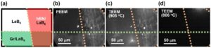
نئی سطح کوٹنگ ٹیکنالوجی مواد کے الیکٹران کے اخراج میں سات گنا اضافہ کرتی ہے۔
ماخذ نوڈ: 2649550
ٹائم اسٹیمپ: 12 فرمائے، 2023
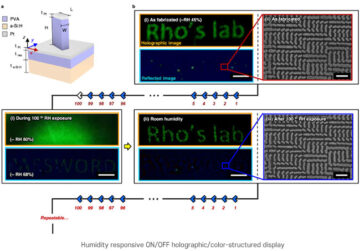
نمی کے جواب میں ہولوگرافک امیجز کے لیے نینو پرنٹنگ تکنیک
ماخذ نوڈ: 1786485
ٹائم اسٹیمپ: دسمبر 26، 2022
دنیا کا اعلیٰ ترین کارکردگی والا ماحول دوست کوانٹم ڈاٹ فوٹو سینسر جس میں کسی بیرونی طاقت کے منبع کی ضرورت نہیں ہے۔
ماخذ نوڈ: 3001715
ٹائم اسٹیمپ: دسمبر 8، 2023