05. Juni 2023 (Nanowerk-Neuigkeiten) Krebschirurgen könnten dank neuer Bildgebungsmittel, die mehrere Biomarker gleichzeitig beleuchten können, bald einen umfassenderen Überblick über Tumore während der Operation erhalten, berichten Forscher der University of Illinois Urbana-Champaign. Die fluoreszierenden Nanopartikel, die in die Membranen roter Blutkörperchen eingewickelt sind, zielen besser auf Tumore ab als derzeit klinisch zugelassene Farbstoffe und können als Reaktion auf nur einen Operationslichtstrahl zwei unterschiedliche Signale aussenden, eine Funktion, die Ärzten helfen könnte, Tumorgrenzen zu unterscheiden und metastasierende Krebsarten zu identifizieren . Die Bildgebungsmittel können mit bioinspirierten Kameras kombiniert werden, die die Forscher zuvor für die Echtzeitdiagnose während der Operation entwickelt haben, sagte Forschungsgruppenleiter Viktor Gruev, Professor für Elektro- und Computertechnik in Illinois. In einer neuen Studie in der Zeitschrift ACS Nano ("Cell-membrane coated nanoparticles for tumor delineation and qualitative estimation of cancer biomarkers at single wavelength excitation in murine and phantom models") demonstrierten die Forscher ihre neuen Dual-Signal-Nanopartikel in Tumorphantomen – 3D-Modellen, die die Merkmale von Tumoren und ihrer Umgebung nachahmen – und in lebenden Mäusen.
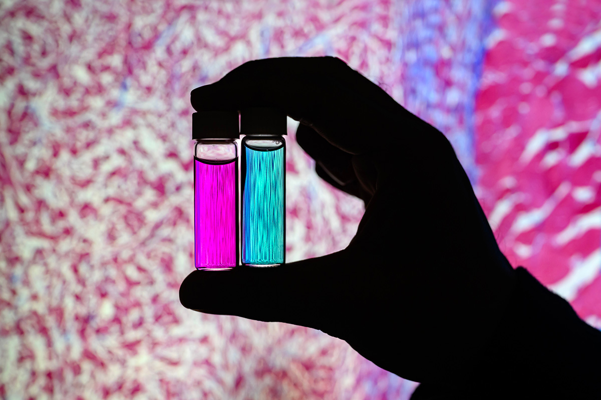 Researcher Indrajit Srivastava holds solutions of nanoparticles that can target two cancer biomarkers, giving off two distinct signals when lit by one fluorescent wavelength. This could give surgeons a more complete picture of a tumor and guide operating-room decisions. In the background is a microscopic scan of a tissue sample. (Image: Fred Zwicky)
“If you want to find all the cancer, imaging one biomarker is not enough. It could miss some tumors. If you introduce a second or a third biomarker, the likelihood of removing all cancer cells increases, and the likelihood of a better outcome for the patients increases.” said Gruev, who also is a professor in the Carle Illinois College of Medicine. “Multiple-targeted drugs and imaging agents are a recent trend, and our group is driving the trend hard because we have the camera technology that can image multiple signals at once.”
Traditionally, a surgeon removes a tumor and sends it to a pathologist for assessment, a process that can take hours to days, said Illinois postdoctoral researcher Indrajit Srivastava, the first author of the paper. As research has moved toward real-time diagnostics, several challenges have prevented wide application: Many tumor-targeted imaging agents only minimally reach their tumor targets, instead being quickly cleared from the blood stream and accumulating in the liver, Srivastava said.
“A few people before us have used nanoparticles coated with red blood cells and found they circulate longer – a few days. We saw the same thing in our mice: The membrane-coated nanoparticles circulated longer in blood, with reduced uptake in the liver. Because they were circulating longer, more of the imaging agents accumulated in the tumors, giving us a stronger fluorescent signal,” Srivastava said.
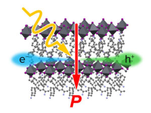
An artist’s rendering of membrane-wrapped nanoparticles binding to markers on a cancer cell and emitting colored light.
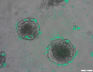
The two biomarkers targeted by the new imaging agents include one that is prevalent in early cancer and one that is prevalent in late-stage cancer, which is more likely to be metastatic. The researchers found that the probes were effective at distinguishing cancerous tissue from healthy tissue, as well as distinguishing the two signals from each other.
“This is appealing for surgical application, as it could help determine where exactly to make the cut. Having multiple signals gives a more overall picture of the tumor. And it could tell a surgeon, ‘This may be metastatic, you may want to be more aggressive in your removal.’” Srivastava said.
Only needing one wavelength of laser light to elicit multiple signals is another benefit for surgical applications, as it makes the instrumentation much more compact than those requiring multiple lasers for each needed wavelength, Gruev said.
The researchers plan to develop more tumor-imaging agents that target multiple markers, and to move forward with further preclinical and clinical studies using their dual-signal dyes with surgical goggles they have developed.
“In this battle for ensuring we remove all the cancer cells during surgery, we need investments both in the imaging camera technology and in the tumor targeting agents,” Gruev said.
Researcher Indrajit Srivastava holds solutions of nanoparticles that can target two cancer biomarkers, giving off two distinct signals when lit by one fluorescent wavelength. This could give surgeons a more complete picture of a tumor and guide operating-room decisions. In the background is a microscopic scan of a tissue sample. (Image: Fred Zwicky)
“If you want to find all the cancer, imaging one biomarker is not enough. It could miss some tumors. If you introduce a second or a third biomarker, the likelihood of removing all cancer cells increases, and the likelihood of a better outcome for the patients increases.” said Gruev, who also is a professor in the Carle Illinois College of Medicine. “Multiple-targeted drugs and imaging agents are a recent trend, and our group is driving the trend hard because we have the camera technology that can image multiple signals at once.”
Traditionally, a surgeon removes a tumor and sends it to a pathologist for assessment, a process that can take hours to days, said Illinois postdoctoral researcher Indrajit Srivastava, the first author of the paper. As research has moved toward real-time diagnostics, several challenges have prevented wide application: Many tumor-targeted imaging agents only minimally reach their tumor targets, instead being quickly cleared from the blood stream and accumulating in the liver, Srivastava said.
“A few people before us have used nanoparticles coated with red blood cells and found they circulate longer – a few days. We saw the same thing in our mice: The membrane-coated nanoparticles circulated longer in blood, with reduced uptake in the liver. Because they were circulating longer, more of the imaging agents accumulated in the tumors, giving us a stronger fluorescent signal,” Srivastava said.
An artist’s rendering of membrane-wrapped nanoparticles binding to markers on a cancer cell and emitting colored light.
The two biomarkers targeted by the new imaging agents include one that is prevalent in early cancer and one that is prevalent in late-stage cancer, which is more likely to be metastatic. The researchers found that the probes were effective at distinguishing cancerous tissue from healthy tissue, as well as distinguishing the two signals from each other.
“This is appealing for surgical application, as it could help determine where exactly to make the cut. Having multiple signals gives a more overall picture of the tumor. And it could tell a surgeon, ‘This may be metastatic, you may want to be more aggressive in your removal.’” Srivastava said.
Only needing one wavelength of laser light to elicit multiple signals is another benefit for surgical applications, as it makes the instrumentation much more compact than those requiring multiple lasers for each needed wavelength, Gruev said.
The researchers plan to develop more tumor-imaging agents that target multiple markers, and to move forward with further preclinical and clinical studies using their dual-signal dyes with surgical goggles they have developed.
“In this battle for ensuring we remove all the cancer cells during surgery, we need investments both in the imaging camera technology and in the tumor targeting agents,” Gruev said.
- SEO-gestützte Content- und PR-Distribution. Holen Sie sich noch heute Verstärkung.
- PlatoAiStream. Web3-Datenintelligenz. Wissen verstärkt. Hier zugreifen.
- Die Zukunft prägen mit Adryenn Ashley. Hier zugreifen.
- Kaufen und verkaufen Sie Anteile an PRE-IPO-Unternehmen mit PREIPO®. Hier zugreifen.
- Quelle: https://www.nanowerk.com/news2/biotech/newsid=63117.php
- :hast
- :Ist
- :nicht
- :Wo
- $UP
- 10
- 12
- 3d
- 7
- 8
- 9
- a
- Angesammelt
- Agenten
- aggressiv
- Alle
- ebenfalls
- an
- und
- Ein anderer
- ansprechend
- Anwendung
- Anwendungen
- Ansatz
- genehmigt
- SIND
- AS
- Bewertung
- At
- Autor
- Hintergrund
- Schlacht
- BE
- Strahl
- weil
- Bevor
- Sein
- Nutzen
- Besser
- Bindung
- Biomarker
- Blut
- Grenzen
- beide
- by
- Kamera
- Kameras
- CAN
- Krebs
- Krebszellen
- Die Zellen
- Center
- Herausforderungen
- zirkulierende
- Klinische
- klinische Versuche
- näher
- Hochschule
- kombiniert
- abschließen
- Computer
- Informationstechnik
- könnte
- Strom
- Schneiden
- Datum
- Tage
- Entscheidungen
- weisen nach, dass
- Bestimmen
- entwickeln
- entwickelt
- deutlich
- unterscheiden
- Ärzte
- Fahren
- Drogen
- im
- jeder
- Früh
- Effektiv
- Entwicklung
- genug
- Gewährleistung
- genau
- Merkmal
- Eigenschaften
- wenige
- Finden Sie
- Vorname
- Aussichten für
- vorwärts
- gefunden
- für
- weiter
- bekommen
- ABSICHT
- gibt
- Unterstützung
- Gruppe an
- Guide
- Pflege
- hart
- Haben
- mit
- gesund
- Hilfe
- Unternehmen
- Halten
- hält
- ganzheitliche
- STUNDEN
- HTTPS
- identifizieren
- if
- Illinois
- beleuchten
- Image
- Imaging
- in
- das
- Steigert
- beantragen müssen
- einführen
- Investments
- IT
- Zeitschrift
- jpg
- nur
- nur einer
- laser
- -Laser
- Führer
- !
- wahrscheinlich
- leben
- Leber
- länger
- um
- MACHT
- viele
- Kann..
- Medizin
- Mäuse
- Mitte
- für
- mehr
- schlauer bewegen
- vorwärts gehen
- viel
- mehrere
- Need
- erforderlich
- benötigen
- Neu
- of
- WOW!
- on
- einmal
- EINEM
- einzige
- or
- Andere
- UNSERE
- Ergebnis
- Gesamt-
- Papier
- Patienten
- Personen
- Phantom
- ein Bild
- Plan
- Plato
- Datenintelligenz von Plato
- PlatoData
- vorherrschend
- vorher
- Prozessdefinierung
- Professor
- qualitativ
- schnell
- erreichen
- Echtzeit
- realisieren
- kürzlich
- Rot
- Reduziert
- Entfernung
- entfernen
- Entfernen
- Rendering
- berichten
- Forschungsprojekte
- Forschungsgruppe
- Forscher
- Forscher
- Antwort
- Said
- gleich
- Scan
- Zweite
- sendet
- mehrere
- Signal
- Signale
- Single
- Lösungen
- einige
- Bald
- Strom
- stärker
- Es wurden Studien
- Studie
- Chirurgie
- chirurgisch
- Nehmen
- Einnahme
- Target
- gezielt
- Targeting
- Ziele
- Technologie
- erzählen
- als
- dank
- zur Verbesserung der Gesundheitsgerechtigkeit
- Das
- ihr
- vom Nutzer definierten
- Ding
- Dritte
- fehlen uns die Worte.
- diejenigen
- zu
- gegenüber
- traditionell
- Trend
- Studien
- XNUMX
- Universität
- us
- benutzt
- Verwendung von
- Anzeigen
- wollen
- we
- GUT
- waren
- wann
- welche
- WHO
- breit
- mit
- Arbeiten
- Eingehüllt
- U
- Ihr
- Zephyrnet
Mehr von Nanowerk
Ein nanoskaliger Nickel-Platin-Kern mit einer Platinhülle spaltet Sauerstoffmoleküle in nützliche Ionen
Quellknoten: 2788122
Zeitstempel: 27. Juli 2023
Die Entdeckung der DNA-Reparatur könnte die Biotechnologie verbessern (mit Video)
Quellknoten: 1987323
Zeitstempel: 2. März 2023
Ein effektiverer Versuchsaufbau, um eine Zelle in einen neuen Zustand zu versetzen
Quellknoten: 2914286
Zeitstempel: 2. Oktober 2023

Forscher entwickeln Bakterien, die Tumor-DNA erkennen können
Quellknoten: 2820250
Zeitstempel: 12. August 2023

Origami-basierte Integration von Robotern, die wahrnehmen, entscheiden und reagieren
Quellknoten: 2565107
Zeitstempel: 4. April 2023
Wir beleuchten den Ursprung des photovoltaischen Effekts in organisch-anorganischen Perowskiten
Quellknoten: 3036090
Zeitstempel: 26. Dezember 2023
Ein Quantensprung in der mechanischen Oszillatortechnologie
Quellknoten: 2817575
Zeitstempel: 11. August 2023
Neue Oberflächenbeschichtungstechnologie erhöht die Elektronenemission von Materialien um das Siebenfache
Quellknoten: 2649550
Zeitstempel: 12. Mai 2023
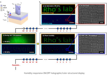
Nanoimprinting-Technik für feuchtigkeitsempfindliche holografische Bilder
Quellknoten: 1786485
Zeitstempel: 26. Dezember 2022
Erste transiente elektronische Bandage beschleunigt die Heilung um 30 %
Quellknoten: 1972187
Zeitstempel: 22. Februar 2023
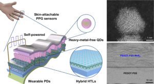
Der weltweit leistungsstärkste umweltfreundliche Quantenpunkt-Fotosensor, für den keine externe Stromquelle erforderlich ist
Quellknoten: 3001715
Zeitstempel: 8. Dezember 2023