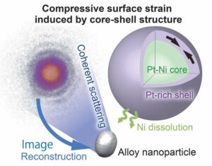
05年2023月XNUMX日(Nanowerk新闻) 伊利诺伊大学厄巴纳-香槟分校的研究人员报告说,由于新的成像剂可以同时照亮多个生物标志物,癌症外科医生可能很快就能在手术过程中对肿瘤有更全面的了解。 包裹在红细胞膜中的荧光纳米粒子比目前临床批准的染料更好地瞄准肿瘤,并且只需一束手术光束即可发出两种不同的信号,这一功能可以帮助医生区分肿瘤边界并识别转移性癌症。 研究小组负责人、伊利诺伊州电气与计算机工程教授维克托·格鲁耶夫(Viktor Gruev)表示,这些显像剂可以与研究人员之前开发的仿生相机相结合,用于手术期间的实时诊断。 在该杂志的一项新研究中 ACS纳米 ("Cell-membrane coated nanoparticles for tumor delineation and qualitative estimation of cancer biomarkers at single wavelength excitation in murine and phantom models"),研究人员在肿瘤模型(模仿肿瘤及其周围环境的 3D 模型)和活体小鼠中展示了他们的新型双信号纳米粒子。
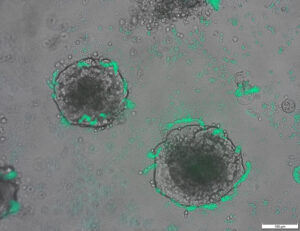
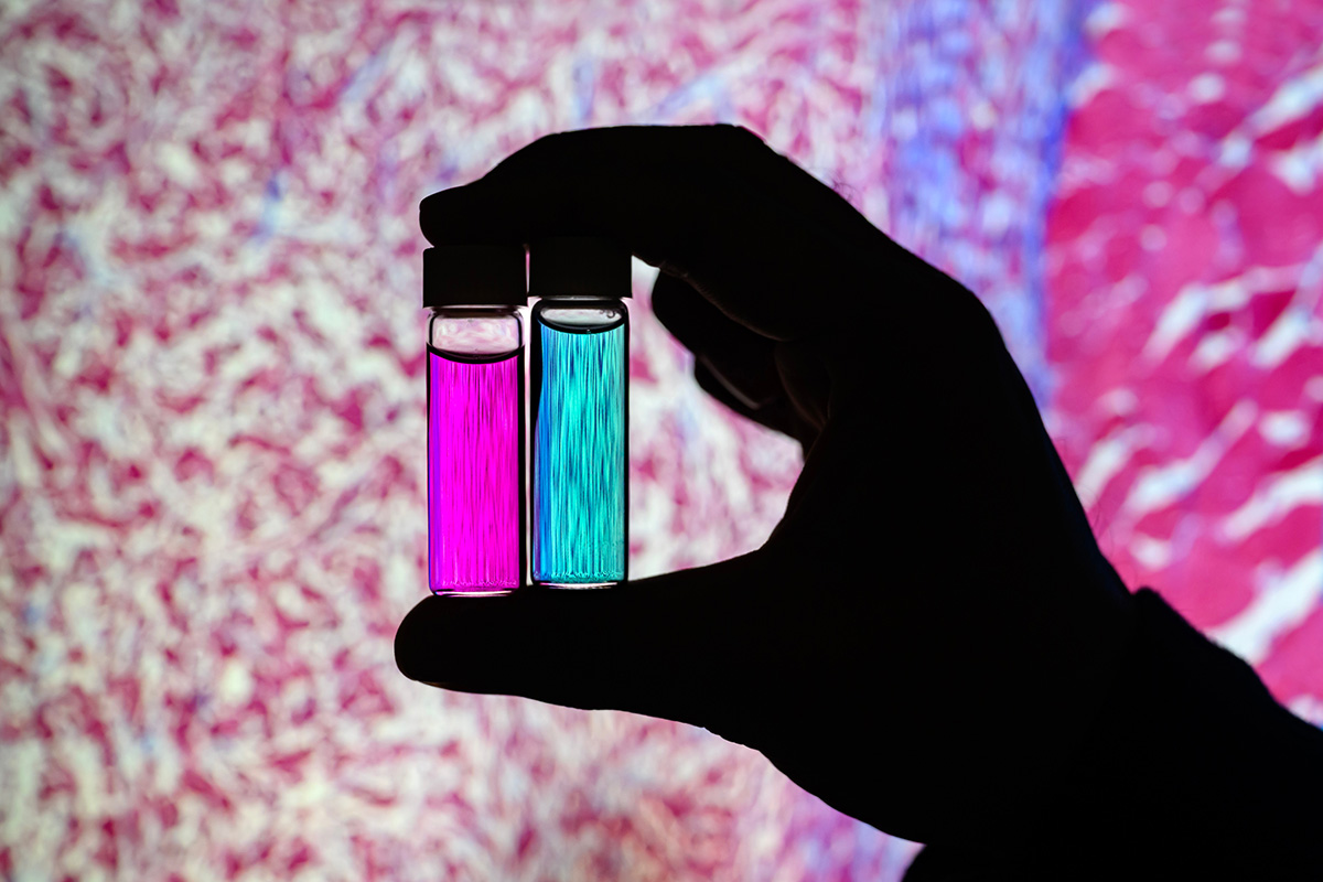 Researcher Indrajit Srivastava holds solutions of nanoparticles that can target two cancer biomarkers, giving off two distinct signals when lit by one fluorescent wavelength. This could give surgeons a more complete picture of a tumor and guide operating-room decisions. In the background is a microscopic scan of a tissue sample. (Image: Fred Zwicky)
“If you want to find all the cancer, imaging one biomarker is not enough. It could miss some tumors. If you introduce a second or a third biomarker, the likelihood of removing all cancer cells increases, and the likelihood of a better outcome for the patients increases.” said Gruev, who also is a professor in the Carle Illinois College of Medicine. “Multiple-targeted drugs and imaging agents are a recent trend, and our group is driving the trend hard because we have the camera technology that can image multiple signals at once.”
Traditionally, a surgeon removes a tumor and sends it to a pathologist for assessment, a process that can take hours to days, said Illinois postdoctoral researcher Indrajit Srivastava, the first author of the paper. As research has moved toward real-time diagnostics, several challenges have prevented wide application: Many tumor-targeted imaging agents only minimally reach their tumor targets, instead being quickly cleared from the blood stream and accumulating in the liver, Srivastava said.
“A few people before us have used nanoparticles coated with red blood cells and found they circulate longer – a few days. We saw the same thing in our mice: The membrane-coated nanoparticles circulated longer in blood, with reduced uptake in the liver. Because they were circulating longer, more of the imaging agents accumulated in the tumors, giving us a stronger fluorescent signal,” Srivastava said.
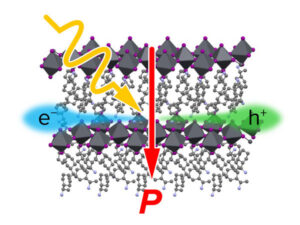
An artist’s rendering of membrane-wrapped nanoparticles binding to markers on a cancer cell and emitting colored light.
The two biomarkers targeted by the new imaging agents include one that is prevalent in early cancer and one that is prevalent in late-stage cancer, which is more likely to be metastatic. The researchers found that the probes were effective at distinguishing cancerous tissue from healthy tissue, as well as distinguishing the two signals from each other.
“This is appealing for surgical application, as it could help determine where exactly to make the cut. Having multiple signals gives a more overall picture of the tumor. And it could tell a surgeon, ‘This may be metastatic, you may want to be more aggressive in your removal.’” Srivastava said.
Only needing one wavelength of laser light to elicit multiple signals is another benefit for surgical applications, as it makes the instrumentation much more compact than those requiring multiple lasers for each needed wavelength, Gruev said.
The researchers plan to develop more tumor-imaging agents that target multiple markers, and to move forward with further preclinical and clinical studies using their dual-signal dyes with surgical goggles they have developed.
“In this battle for ensuring we remove all the cancer cells during surgery, we need investments both in the imaging camera technology and in the tumor targeting agents,” Gruev said.
Researcher Indrajit Srivastava holds solutions of nanoparticles that can target two cancer biomarkers, giving off two distinct signals when lit by one fluorescent wavelength. This could give surgeons a more complete picture of a tumor and guide operating-room decisions. In the background is a microscopic scan of a tissue sample. (Image: Fred Zwicky)
“If you want to find all the cancer, imaging one biomarker is not enough. It could miss some tumors. If you introduce a second or a third biomarker, the likelihood of removing all cancer cells increases, and the likelihood of a better outcome for the patients increases.” said Gruev, who also is a professor in the Carle Illinois College of Medicine. “Multiple-targeted drugs and imaging agents are a recent trend, and our group is driving the trend hard because we have the camera technology that can image multiple signals at once.”
Traditionally, a surgeon removes a tumor and sends it to a pathologist for assessment, a process that can take hours to days, said Illinois postdoctoral researcher Indrajit Srivastava, the first author of the paper. As research has moved toward real-time diagnostics, several challenges have prevented wide application: Many tumor-targeted imaging agents only minimally reach their tumor targets, instead being quickly cleared from the blood stream and accumulating in the liver, Srivastava said.
“A few people before us have used nanoparticles coated with red blood cells and found they circulate longer – a few days. We saw the same thing in our mice: The membrane-coated nanoparticles circulated longer in blood, with reduced uptake in the liver. Because they were circulating longer, more of the imaging agents accumulated in the tumors, giving us a stronger fluorescent signal,” Srivastava said.
An artist’s rendering of membrane-wrapped nanoparticles binding to markers on a cancer cell and emitting colored light.
The two biomarkers targeted by the new imaging agents include one that is prevalent in early cancer and one that is prevalent in late-stage cancer, which is more likely to be metastatic. The researchers found that the probes were effective at distinguishing cancerous tissue from healthy tissue, as well as distinguishing the two signals from each other.
“This is appealing for surgical application, as it could help determine where exactly to make the cut. Having multiple signals gives a more overall picture of the tumor. And it could tell a surgeon, ‘This may be metastatic, you may want to be more aggressive in your removal.’” Srivastava said.
Only needing one wavelength of laser light to elicit multiple signals is another benefit for surgical applications, as it makes the instrumentation much more compact than those requiring multiple lasers for each needed wavelength, Gruev said.
The researchers plan to develop more tumor-imaging agents that target multiple markers, and to move forward with further preclinical and clinical studies using their dual-signal dyes with surgical goggles they have developed.
“In this battle for ensuring we remove all the cancer cells during surgery, we need investments both in the imaging camera technology and in the tumor targeting agents,” Gruev said.
- SEO 支持的内容和 PR 分发。 今天得到放大。
- 柏拉图爱流。 Web3 数据智能。 知识放大。 访问这里。
- 与 Adryenn Ashley 一起铸造未来。 访问这里。
- 使用 PREIPO® 买卖 PRE-IPO 公司的股票。 访问这里。
- Sumber: https://www.nanowerk.com/news2/biotech/newsid=63117.php
- :具有
- :是
- :不是
- :在哪里
- $UP
- 10
- 12
- 3d
- 7
- 8
- 9
- a
- 积累
- 中介代理
- 侵略性
- 所有类型
- 还
- an
- 和
- 另一个
- 吸引人的
- 应用领域
- 应用领域
- 的途径
- 批准
- 保健
- AS
- 评定
- At
- 作者
- 背景
- 战斗
- BE
- 光束
- 因为
- before
- 作为
- 得益
- 更好
- 捆绑
- 生物标记物
- 血液
- 边界
- 都
- by
- 相机
- 相机
- CAN
- 癌症预防
- 癌细胞
- 细胞
- Center
- 挑战
- 循环
- 临床资料
- 临床试验
- 接近
- 学院
- 结合
- 完成
- 一台
- 计算机工程
- 可以
- 电流
- 切
- 日期
- 一年中的
- 决定
- 证明
- 确定
- 开发
- 发达
- 不同
- 区分
- 医生
- 驾驶
- 毒品
- ,我们将参加
- 每
- 早
- 有效
- 工程师
- 更多
- 保证
- 究竟
- 专栏
- 特征
- 少数
- 找到最适合您的地方
- 姓氏:
- 针对
- 向前
- 发现
- 止
- 进一步
- 越来越
- 给
- 给
- 给予
- 团队
- 指南
- 手
- 硬
- 有
- 有
- 健康
- 帮助
- 帮助
- 保持
- 持有
- 整体
- HOURS
- HTTPS
- 鉴定
- if
- 伊利诺伊州
- 照亮
- 图片
- 同步成像
- in
- 包括
- 增加
- 代替
- 介绍
- 投资
- IT
- 日志
- JPG
- 只是
- 只有一个
- 激光器是如何工作的
- 激光器
- 领导者
- 光
- 容易
- 生活
- 肝
- 不再
- 使
- 制作
- 许多
- 可能..
- 药物
- 小鼠
- 中间
- 模型
- 更多
- 移动
- 前进
- 许多
- 多
- 需求
- 打印车票
- 需要
- 全新
- of
- 折扣
- on
- 一旦
- 一
- 仅由
- or
- 其他名称
- 我们的
- 成果
- 最划算
- 纸类
- 患者
- 员工
- 模体
- 图片
- 计划
- 柏拉图
- 柏拉图数据智能
- 柏拉图数据
- 流行
- 先前
- 过程
- 教授
- 定性
- 很快
- 达到
- 实时的
- 实现
- 最近
- 红色
- 减少
- 切除
- 去掉
- 删除
- 翻译
- 报告
- 研究
- 研究小组
- 研究员
- 研究人员
- 响应
- 说
- 同
- 浏览
- 其次
- 发送
- 几个
- 信号
- 信号
- 单
- 解决方案
- 一些
- 不久
- 流
- 强
- 研究
- 学习
- 手术
- 外科
- 采取
- 服用
- 目标
- 针对
- 瞄准
- 目标
- 专业技术
- 展示
- 比
- 谢谢
- 这
- 其
- 他们
- 事
- 第三
- Free Introduction
- 那些
- 至
- 对于
- 传统
- 趋势
- 试验
- 二
- 大学
- us
- 用过的
- 运用
- 查看
- 想
- we
- 井
- 为
- ,尤其是
- 这
- WHO
- 宽
- 工作
- 包裹
- 您
- 您一站式解决方案
- 和风网