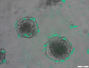
05 มิ.ย. 2023 (ข่าวนาโนเวิร์ค) ศัลยแพทย์มะเร็งอาจมีมุมมองที่สมบูรณ์ยิ่งขึ้นของเนื้องอกในระหว่างการผ่าตัดในไม่ช้า เนื่องจากมีสารสร้างภาพใหม่ที่สามารถส่องตัวบ่งชี้ทางชีวภาพหลายตัวในคราวเดียว นักวิจัยจากมหาวิทยาลัยอิลลินอยส์ เออร์บานา-แชมเพน รายงาน อนุภาคนาโนเรืองแสงที่ห่อหุ้มอยู่ในเยื่อหุ้มเซลล์เม็ดเลือดแดง มุ่งเป้าไปที่เนื้องอกได้ดีกว่าสีย้อมที่ได้รับการรับรองทางคลินิกในปัจจุบัน และสามารถส่งสัญญาณที่แตกต่างกันสองสัญญาณเพื่อตอบสนองต่อลำแสงผ่าตัดเพียงลำแสงเดียว ซึ่งเป็นคุณสมบัติที่ช่วยให้แพทย์แยกแยะขอบเขตของเนื้องอกและระบุมะเร็งระยะลุกลามได้ . สารสร้างภาพสามารถใช้ร่วมกับกล้องที่ได้รับแรงบันดาลใจจากชีวภาพ ซึ่งก่อนหน้านี้นักวิจัยได้พัฒนาขึ้นเพื่อการวินิจฉัยแบบเรียลไทม์ระหว่างการผ่าตัด หัวหน้ากลุ่มวิจัย Viktor Gruev ศาสตราจารย์ด้านวิศวกรรมไฟฟ้าและคอมพิวเตอร์ในรัฐอิลลินอยส์ กล่าว ในการศึกษาใหม่ในวารสาร ACS Nano ("Cell-membrane coated nanoparticles for tumor delineation and qualitative estimation of cancer biomarkers at single wavelength excitation in murine and phantom models") นักวิจัยได้สาธิตอนุภาคนาโนสัญญาณคู่แบบใหม่ในแบบจำลอง 3 มิติที่เลียนแบบลักษณะของเนื้องอกและสภาพแวดล้อม และในหนูที่มีชีวิต
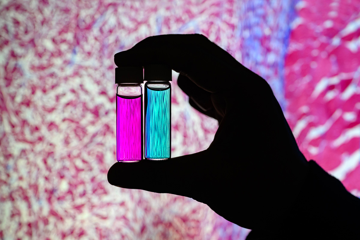 Researcher Indrajit Srivastava holds solutions of nanoparticles that can target two cancer biomarkers, giving off two distinct signals when lit by one fluorescent wavelength. This could give surgeons a more complete picture of a tumor and guide operating-room decisions. In the background is a microscopic scan of a tissue sample. (Image: Fred Zwicky)
“If you want to find all the cancer, imaging one biomarker is not enough. It could miss some tumors. If you introduce a second or a third biomarker, the likelihood of removing all cancer cells increases, and the likelihood of a better outcome for the patients increases.” said Gruev, who also is a professor in the Carle Illinois College of Medicine. “Multiple-targeted drugs and imaging agents are a recent trend, and our group is driving the trend hard because we have the camera technology that can image multiple signals at once.”
Traditionally, a surgeon removes a tumor and sends it to a pathologist for assessment, a process that can take hours to days, said Illinois postdoctoral researcher Indrajit Srivastava, the first author of the paper. As research has moved toward real-time diagnostics, several challenges have prevented wide application: Many tumor-targeted imaging agents only minimally reach their tumor targets, instead being quickly cleared from the blood stream and accumulating in the liver, Srivastava said.
“A few people before us have used nanoparticles coated with red blood cells and found they circulate longer – a few days. We saw the same thing in our mice: The membrane-coated nanoparticles circulated longer in blood, with reduced uptake in the liver. Because they were circulating longer, more of the imaging agents accumulated in the tumors, giving us a stronger fluorescent signal,” Srivastava said.
An artist’s rendering of membrane-wrapped nanoparticles binding to markers on a cancer cell and emitting colored light.
The two biomarkers targeted by the new imaging agents include one that is prevalent in early cancer and one that is prevalent in late-stage cancer, which is more likely to be metastatic. The researchers found that the probes were effective at distinguishing cancerous tissue from healthy tissue, as well as distinguishing the two signals from each other.
“This is appealing for surgical application, as it could help determine where exactly to make the cut. Having multiple signals gives a more overall picture of the tumor. And it could tell a surgeon, ‘This may be metastatic, you may want to be more aggressive in your removal.’” Srivastava said.
Only needing one wavelength of laser light to elicit multiple signals is another benefit for surgical applications, as it makes the instrumentation much more compact than those requiring multiple lasers for each needed wavelength, Gruev said.
The researchers plan to develop more tumor-imaging agents that target multiple markers, and to move forward with further preclinical and clinical studies using their dual-signal dyes with surgical goggles they have developed.
“In this battle for ensuring we remove all the cancer cells during surgery, we need investments both in the imaging camera technology and in the tumor targeting agents,” Gruev said.
Researcher Indrajit Srivastava holds solutions of nanoparticles that can target two cancer biomarkers, giving off two distinct signals when lit by one fluorescent wavelength. This could give surgeons a more complete picture of a tumor and guide operating-room decisions. In the background is a microscopic scan of a tissue sample. (Image: Fred Zwicky)
“If you want to find all the cancer, imaging one biomarker is not enough. It could miss some tumors. If you introduce a second or a third biomarker, the likelihood of removing all cancer cells increases, and the likelihood of a better outcome for the patients increases.” said Gruev, who also is a professor in the Carle Illinois College of Medicine. “Multiple-targeted drugs and imaging agents are a recent trend, and our group is driving the trend hard because we have the camera technology that can image multiple signals at once.”
Traditionally, a surgeon removes a tumor and sends it to a pathologist for assessment, a process that can take hours to days, said Illinois postdoctoral researcher Indrajit Srivastava, the first author of the paper. As research has moved toward real-time diagnostics, several challenges have prevented wide application: Many tumor-targeted imaging agents only minimally reach their tumor targets, instead being quickly cleared from the blood stream and accumulating in the liver, Srivastava said.
“A few people before us have used nanoparticles coated with red blood cells and found they circulate longer – a few days. We saw the same thing in our mice: The membrane-coated nanoparticles circulated longer in blood, with reduced uptake in the liver. Because they were circulating longer, more of the imaging agents accumulated in the tumors, giving us a stronger fluorescent signal,” Srivastava said.
An artist’s rendering of membrane-wrapped nanoparticles binding to markers on a cancer cell and emitting colored light.
The two biomarkers targeted by the new imaging agents include one that is prevalent in early cancer and one that is prevalent in late-stage cancer, which is more likely to be metastatic. The researchers found that the probes were effective at distinguishing cancerous tissue from healthy tissue, as well as distinguishing the two signals from each other.
“This is appealing for surgical application, as it could help determine where exactly to make the cut. Having multiple signals gives a more overall picture of the tumor. And it could tell a surgeon, ‘This may be metastatic, you may want to be more aggressive in your removal.’” Srivastava said.
Only needing one wavelength of laser light to elicit multiple signals is another benefit for surgical applications, as it makes the instrumentation much more compact than those requiring multiple lasers for each needed wavelength, Gruev said.
The researchers plan to develop more tumor-imaging agents that target multiple markers, and to move forward with further preclinical and clinical studies using their dual-signal dyes with surgical goggles they have developed.
“In this battle for ensuring we remove all the cancer cells during surgery, we need investments both in the imaging camera technology and in the tumor targeting agents,” Gruev said.
- เนื้อหาที่ขับเคลื่อนด้วย SEO และการเผยแพร่ประชาสัมพันธ์ รับการขยายวันนี้
- เพลโตไอสตรีม. ข้อมูลอัจฉริยะ Web3 ขยายความรู้ เข้าถึงได้ที่นี่.
- การสร้างอนาคตโดย Adryenn Ashley เข้าถึงได้ที่นี่.
- ซื้อและขายหุ้นในบริษัท PRE-IPO ด้วย PREIPO® เข้าถึงได้ที่นี่.
- ที่มา: https://www.nanowerk.com/news2/biotech/newsid=63117.php
- :มี
- :เป็น
- :ไม่
- :ที่ไหน
- $ ขึ้น
- 10
- 12
- 3d
- 7
- 8
- 9
- a
- สะสม
- ตัวแทน
- ก้าวร้าว
- ทั้งหมด
- ด้วย
- an
- และ
- อื่น
- อุทธรณ์
- การใช้งาน
- การใช้งาน
- เข้าใกล้
- ได้รับการอนุมัติ
- เป็น
- AS
- การประเมินผล
- At
- ผู้เขียน
- พื้นหลัง
- การต่อสู้
- BE
- คาน
- เพราะ
- ก่อน
- กำลัง
- ประโยชน์
- ดีกว่า
- ผูกพัน
- biomarker
- เลือด
- พรมแดน
- ทั้งสอง
- by
- ห้อง
- กล้อง
- CAN
- โรคมะเร็ง
- เซลล์มะเร็ง
- เซลล์
- ศูนย์
- ความท้าทาย
- หมุนเวียน
- คลินิก
- การทดลองทางคลินิก
- ใกล้ชิด
- วิทยาลัย
- รวม
- สมบูรณ์
- คอมพิวเตอร์
- วิศวกรรมคอมพิวเตอร์
- ได้
- ปัจจุบัน
- ตัด
- วันที่
- วัน
- การตัดสินใจ
- แสดงให้เห็นถึง
- กำหนด
- พัฒนา
- พัฒนา
- แตกต่าง
- เห็นความแตกต่าง
- แพทย์
- การขับขี่
- ยาเสพติด
- ในระหว่าง
- แต่ละ
- ก่อน
- มีประสิทธิภาพ
- ชั้นเยี่ยม
- พอ
- การสร้างความมั่นใจ
- เผง
- ลักษณะ
- คุณสมบัติ
- สองสาม
- หา
- ชื่อจริง
- สำหรับ
- ข้างหน้า
- พบ
- ราคาเริ่มต้นที่
- ต่อไป
- ได้รับ
- ให้
- จะช่วยให้
- ให้
- บัญชีกลุ่ม
- ให้คำแนะนำ
- มือ
- ยาก
- มี
- มี
- แข็งแรง
- ช่วย
- การช่วยเหลือ
- โฮลดิ้ง
- ถือ
- แบบองค์รวม
- ชั่วโมง
- HTTPS
- แยกแยะ
- if
- อิลลินอยส์
- เปล่ง
- ภาพ
- การถ่ายภาพ
- in
- ประกอบด้วย
- เพิ่มขึ้น
- แทน
- แนะนำ
- เงินลงทุน
- IT
- วารสาร
- jpg
- เพียงแค่
- แค่หนึ่ง
- เลเซอร์
- เลเซอร์
- ผู้นำ
- เบา
- น่าจะ
- สด
- ตับ
- อีกต่อไป
- ทำ
- ทำให้
- หลาย
- อาจ..
- ยา
- หนู
- กลาง
- โมเดล
- ข้อมูลเพิ่มเติม
- ย้าย
- เดินหน้าต่อไป
- มาก
- หลาย
- จำเป็นต้อง
- จำเป็น
- ต้อง
- ใหม่
- of
- ปิด
- on
- ครั้งเดียว
- ONE
- เพียง
- or
- อื่นๆ
- ของเรา
- ผล
- ทั้งหมด
- กระดาษ
- ผู้ป่วย
- คน
- ผี
- ภาพ
- แผนการ
- เพลโต
- เพลโตดาต้าอินเทลลิเจนซ์
- เพลโตดาต้า
- เป็นที่แพร่หลาย
- ก่อนหน้านี้
- กระบวนการ
- ศาสตราจารย์
- เชิงคุณภาพ
- อย่างรวดเร็ว
- มาถึง
- เรียลไทม์
- ตระหนักถึง
- เมื่อเร็ว ๆ นี้
- สีแดง
- ลดลง
- การกำจัด
- เอาออก
- ลบ
- การแสดงผล
- รายงาน
- การวิจัย
- กลุ่มวิจัย
- นักวิจัย
- นักวิจัย
- คำตอบ
- กล่าวว่า
- เดียวกัน
- การสแกน
- ที่สอง
- ส่ง
- หลาย
- สัญญาณ
- สัญญาณ
- เดียว
- โซลูชัน
- บาง
- ในไม่ช้า
- กระแส
- แข็งแกร่ง
- การศึกษา
- ศึกษา
- ศัลยกรรม
- ผ่าตัด
- เอา
- การ
- เป้า
- เป้าหมาย
- กำหนดเป้าหมาย
- เป้าหมาย
- เทคโนโลยี
- บอก
- กว่า
- ขอบคุณ
- ที่
- พื้นที่
- ของพวกเขา
- พวกเขา
- สิ่ง
- ที่สาม
- นี้
- เหล่านั้น
- ไปยัง
- ไปทาง
- ตามธรรมเนียม
- เทรนด์
- การทดลอง
- สอง
- มหาวิทยาลัย
- us
- มือสอง
- การใช้
- รายละเอียด
- ต้องการ
- we
- ดี
- คือ
- เมื่อ
- ที่
- WHO
- กว้าง
- กับ
- งาน
- ตะลึง
- เธอ
- ของคุณ
- ลมทะเล
เพิ่มเติมจาก นาโนเวิร์ค
แกนนาโนสเกลนิกเกิล-แพลตตินัมที่มีเปลือกแพลตตินัมจะแตกโมเลกุลออกซิเจนให้เป็นไอออนที่มีประโยชน์
โหนดต้นทาง: 2788122
ประทับเวลา: กรกฎาคม 27, 2023
การค้นพบการซ่อมแซม DNA สามารถปรับปรุงเทคโนโลยีชีวภาพ (พร้อมวิดีโอ)
โหนดต้นทาง: 1987323
ประทับเวลา: Mar 2, 2023
การออกแบบการทดลองที่มีประสิทธิภาพมากขึ้นสำหรับวิศวกรรมเซลล์ให้อยู่ในสถานะใหม่
โหนดต้นทาง: 2914286
ประทับเวลา: ตุลาคม 2, 2023

นักวิจัยออกแบบแบคทีเรียที่สามารถตรวจจับ DNA ของเนื้องอกได้
โหนดต้นทาง: 2820250
ประทับเวลา: สิงหาคม 12, 2023

การผสานการทำงานของหุ่นยนต์แบบ Origami ที่รับรู้ ตัดสินใจ และตอบสนอง
โหนดต้นทาง: 2565107
ประทับเวลา: เมษายน 4, 2023
การฉายแสงเกี่ยวกับต้นกำเนิดของเอฟเฟกต์ไฟฟ้าโซลาร์เซลล์ในเพอร์รอฟสกี้แบบอินทรีย์และอนินทรีย์
โหนดต้นทาง: 3036090
ประทับเวลา: ธันวาคม 26, 2023
เทคโนโลยีการเคลือบพื้นผิวใหม่ช่วยเพิ่มการปล่อยอิเล็กตรอนของวัสดุเจ็ดเท่า
โหนดต้นทาง: 2649550
ประทับเวลา: May 12, 2023
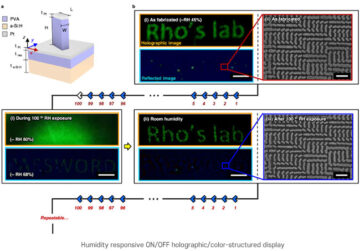
เทคนิคการพิมพ์นาโนสำหรับภาพโฮโลแกรมที่ตอบสนองต่อความชื้น
โหนดต้นทาง: 1786485
ประทับเวลา: ธันวาคม 26, 2022
ผ้าพันแผลอิเล็กทรอนิกส์แบบชั่วคราวชุดแรกจะรักษาเร็วขึ้น 30%
โหนดต้นทาง: 1972187
ประทับเวลา: กุมภาพันธ์ 22, 2023
โฟโตเซ็นเซอร์ควอนตัมดอทประสิทธิภาพสูงที่สุดในโลกที่เป็นมิตรกับสิ่งแวดล้อม โดยไม่ต้องใช้แหล่งพลังงานภายนอก
โหนดต้นทาง: 3001715
ประทับเวลา: ธันวาคม 8, 2023