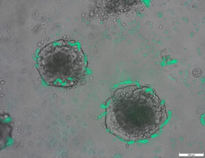
05 iunie 2023 (Știri Nanowerk) Chirurgii de cancer ar putea avea în curând o vedere mai completă a tumorilor în timpul intervenției chirurgicale datorită noilor agenți imagistici care pot ilumina mai mulți biomarkeri simultan, raportează cercetătorii de la Universitatea din Illinois Urbana-Champaign. Nanoparticulele fluorescente, învelite în membranele celulelor roșii din sânge, vizează tumorile mai bine decât coloranții actuali aprobati clinic și pot emite două semnale distincte ca răspuns la un singur fascicul de lumină chirurgicală, o caracteristică care ar putea ajuta medicii să distingă granițele tumorale și să identifice cancerele metastatice. . Agenții de imagistică pot fi combinați cu camere bioinspirate, pe care cercetătorii le-au dezvoltat anterior pentru diagnosticarea în timp real în timpul intervenției chirurgicale, a declarat liderul grupului de cercetare Viktor Gruev, profesor de inginerie electrică și informatică din Illinois. Într-un nou studiu în jurnal ACS Nano ("Cell-membrane coated nanoparticles for tumor delineation and qualitative estimation of cancer biomarkers at single wavelength excitation in murine and phantom models"), cercetătorii și-au demonstrat noile nanoparticule cu semnal dublu în fantomele tumorale - modele 3D care imită caracteristicile tumorilor și ale împrejurimilor lor - și la șoarecii vii.
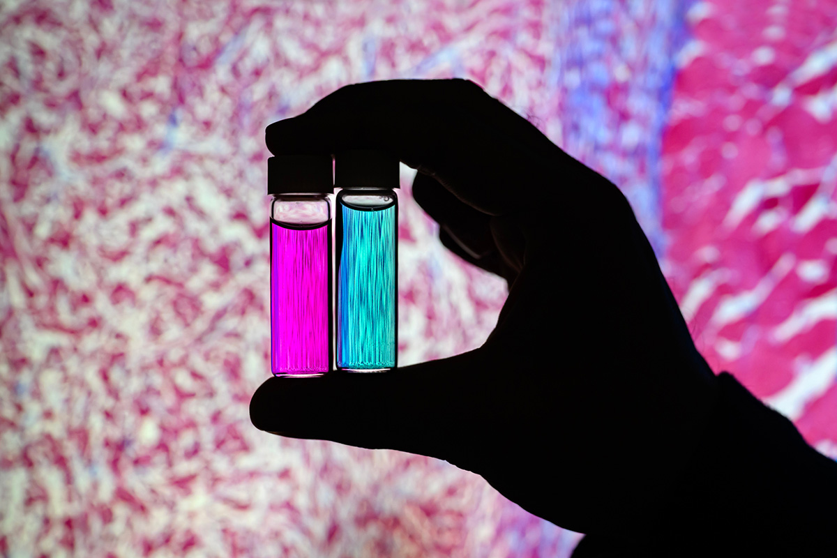 Researcher Indrajit Srivastava holds solutions of nanoparticles that can target two cancer biomarkers, giving off two distinct signals when lit by one fluorescent wavelength. This could give surgeons a more complete picture of a tumor and guide operating-room decisions. In the background is a microscopic scan of a tissue sample. (Image: Fred Zwicky)
“If you want to find all the cancer, imaging one biomarker is not enough. It could miss some tumors. If you introduce a second or a third biomarker, the likelihood of removing all cancer cells increases, and the likelihood of a better outcome for the patients increases.” said Gruev, who also is a professor in the Carle Illinois College of Medicine. “Multiple-targeted drugs and imaging agents are a recent trend, and our group is driving the trend hard because we have the camera technology that can image multiple signals at once.”
Traditionally, a surgeon removes a tumor and sends it to a pathologist for assessment, a process that can take hours to days, said Illinois postdoctoral researcher Indrajit Srivastava, the first author of the paper. As research has moved toward real-time diagnostics, several challenges have prevented wide application: Many tumor-targeted imaging agents only minimally reach their tumor targets, instead being quickly cleared from the blood stream and accumulating in the liver, Srivastava said.
“A few people before us have used nanoparticles coated with red blood cells and found they circulate longer – a few days. We saw the same thing in our mice: The membrane-coated nanoparticles circulated longer in blood, with reduced uptake in the liver. Because they were circulating longer, more of the imaging agents accumulated in the tumors, giving us a stronger fluorescent signal,” Srivastava said.
An artist’s rendering of membrane-wrapped nanoparticles binding to markers on a cancer cell and emitting colored light.
The two biomarkers targeted by the new imaging agents include one that is prevalent in early cancer and one that is prevalent in late-stage cancer, which is more likely to be metastatic. The researchers found that the probes were effective at distinguishing cancerous tissue from healthy tissue, as well as distinguishing the two signals from each other.
“This is appealing for surgical application, as it could help determine where exactly to make the cut. Having multiple signals gives a more overall picture of the tumor. And it could tell a surgeon, ‘This may be metastatic, you may want to be more aggressive in your removal.’” Srivastava said.
Only needing one wavelength of laser light to elicit multiple signals is another benefit for surgical applications, as it makes the instrumentation much more compact than those requiring multiple lasers for each needed wavelength, Gruev said.
The researchers plan to develop more tumor-imaging agents that target multiple markers, and to move forward with further preclinical and clinical studies using their dual-signal dyes with surgical goggles they have developed.
“In this battle for ensuring we remove all the cancer cells during surgery, we need investments both in the imaging camera technology and in the tumor targeting agents,” Gruev said.
Researcher Indrajit Srivastava holds solutions of nanoparticles that can target two cancer biomarkers, giving off two distinct signals when lit by one fluorescent wavelength. This could give surgeons a more complete picture of a tumor and guide operating-room decisions. In the background is a microscopic scan of a tissue sample. (Image: Fred Zwicky)
“If you want to find all the cancer, imaging one biomarker is not enough. It could miss some tumors. If you introduce a second or a third biomarker, the likelihood of removing all cancer cells increases, and the likelihood of a better outcome for the patients increases.” said Gruev, who also is a professor in the Carle Illinois College of Medicine. “Multiple-targeted drugs and imaging agents are a recent trend, and our group is driving the trend hard because we have the camera technology that can image multiple signals at once.”
Traditionally, a surgeon removes a tumor and sends it to a pathologist for assessment, a process that can take hours to days, said Illinois postdoctoral researcher Indrajit Srivastava, the first author of the paper. As research has moved toward real-time diagnostics, several challenges have prevented wide application: Many tumor-targeted imaging agents only minimally reach their tumor targets, instead being quickly cleared from the blood stream and accumulating in the liver, Srivastava said.
“A few people before us have used nanoparticles coated with red blood cells and found they circulate longer – a few days. We saw the same thing in our mice: The membrane-coated nanoparticles circulated longer in blood, with reduced uptake in the liver. Because they were circulating longer, more of the imaging agents accumulated in the tumors, giving us a stronger fluorescent signal,” Srivastava said.
An artist’s rendering of membrane-wrapped nanoparticles binding to markers on a cancer cell and emitting colored light.
The two biomarkers targeted by the new imaging agents include one that is prevalent in early cancer and one that is prevalent in late-stage cancer, which is more likely to be metastatic. The researchers found that the probes were effective at distinguishing cancerous tissue from healthy tissue, as well as distinguishing the two signals from each other.
“This is appealing for surgical application, as it could help determine where exactly to make the cut. Having multiple signals gives a more overall picture of the tumor. And it could tell a surgeon, ‘This may be metastatic, you may want to be more aggressive in your removal.’” Srivastava said.
Only needing one wavelength of laser light to elicit multiple signals is another benefit for surgical applications, as it makes the instrumentation much more compact than those requiring multiple lasers for each needed wavelength, Gruev said.
The researchers plan to develop more tumor-imaging agents that target multiple markers, and to move forward with further preclinical and clinical studies using their dual-signal dyes with surgical goggles they have developed.
“In this battle for ensuring we remove all the cancer cells during surgery, we need investments both in the imaging camera technology and in the tumor targeting agents,” Gruev said.
- Distribuție de conținut bazat pe SEO și PR. Amplifică-te astăzi.
- PlatoAiStream. Web3 Data Intelligence. Cunoștințe amplificate. Accesați Aici.
- Mintând viitorul cu Adryenn Ashley. Accesați Aici.
- Cumpărați și vindeți acțiuni în companii PRE-IPO cu PREIPO®. Accesați Aici.
- Sursa: https://www.nanowerk.com/news2/biotech/newsid=63117.php
- :are
- :este
- :nu
- :Unde
- $UP
- 10
- 12
- 3d
- 7
- 8
- 9
- a
- acumulate
- agenţi
- agresiv
- TOATE
- de asemenea
- an
- și
- O alta
- interesant
- aplicație
- aplicatii
- abordare
- aprobat
- SUNT
- AS
- evaluare
- At
- autor
- fundal
- Luptă
- BE
- Grindă
- deoarece
- înainte
- fiind
- beneficia
- Mai bine
- legare
- biomarker
- sânge
- frontierelor
- atât
- by
- aparat foto
- camere video
- CAN
- Rac
- Celule canceroase
- Celule
- Centru
- provocări
- circulant
- clinic
- studii clinice
- mai aproape
- Colegiu
- combinate
- Completă
- calculator
- Inginerie calculator
- ar putea
- Curent
- Tăiat
- Data
- Zi
- Deciziile
- demonstrat
- Determina
- dezvolta
- dezvoltat
- distinct
- distinge
- Medici
- conducere
- Droguri
- în timpul
- fiecare
- Devreme
- Eficace
- Inginerie
- suficient de
- asigurare
- exact
- Caracteristică
- DESCRIERE
- puțini
- Găsi
- First
- Pentru
- Înainte
- găsit
- din
- mai mult
- obtinerea
- Da
- oferă
- Oferirea
- grup
- ghida
- mână
- Greu
- Avea
- având în
- sănătos
- ajutor
- ajutor
- deținere
- deține
- holistică
- ORE
- HTTPS
- identifica
- if
- Illinois
- ilumina
- imagine
- Imaging
- in
- include
- Creșteri
- in schimb
- introduce
- Investiții
- IT
- jurnal
- jpg
- doar
- doar unul
- cu laser
- lasere
- lider
- ușoară
- Probabil
- trăi
- Ficat
- mai lung
- face
- FACE
- multe
- Mai..
- medicină
- soareci
- De mijloc
- Modele
- mai mult
- muta
- mergi inainte
- mult
- multiplu
- Nevoie
- necesar
- au nevoie
- Nou
- of
- de pe
- on
- dată
- ONE
- afară
- or
- Altele
- al nostru
- Rezultat
- global
- Hârtie
- pacientes
- oameni
- fantomă
- imagine
- plan
- Plato
- Informații despre date Platon
- PlatoData
- prevalent
- în prealabil
- proces
- Profesor
- calitativ
- repede
- ajunge
- în timp real
- realiza
- recent
- Roșu
- Redus
- îndepărtare
- scoate
- eliminarea
- tencuială
- raportează
- cercetare
- grup de cercetare
- cercetător
- cercetători
- răspuns
- Said
- acelaşi
- scanare
- Al doilea
- trimite
- câteva
- Semnal
- semnalele
- singur
- soluţii
- unele
- Curând
- curent
- puternic
- studiu
- Studiu
- Intervenție Chirurgicală
- chirurgical
- Lua
- luare
- Ţintă
- vizate
- direcționare
- obiective
- Tehnologia
- spune
- decât
- Mulțumiri
- acea
- lor
- ei
- lucru
- Al treilea
- acest
- aceste
- la
- spre
- tradiţional
- tendință
- studii
- Două
- universitate
- us
- utilizat
- folosind
- Vizualizare
- vrea
- we
- BINE
- au fost
- cand
- care
- OMS
- larg
- cu
- Apartamente
- Înfășurat
- tu
- Ta
- zephyrnet
Mai mult de la Nanowerk
Un miez la scară nanometrică de nichel-platină cu o înveliș de platină sparge moleculele de oxigen în ioni utili
Nodul sursă: 2788122
Timestamp-ul: Iulie 27, 2023
Descoperirea reparației ADN-ului ar putea îmbunătăți biotehnologia (cu video)
Nodul sursă: 1987323
Timestamp-ul: Mar 2, 2023
Un proiect experimental mai eficient pentru proiectarea unei celule într-o stare nouă
Nodul sursă: 2914286
Timestamp-ul: Octombrie 2, 2023

Cercetătorii creează bacterii care pot detecta ADN-ul tumorii
Nodul sursă: 2820250
Timestamp-ul: August 12, 2023
Integrarea bazată pe origami a roboților care simt, decid și răspund
Nodul sursă: 2565107
Timestamp-ul: Aprilie 4, 2023
Aruncarea lumină asupra originii efectului fotovoltaic în perovskiții organic-anorganici
Nodul sursă: 3036090
Timestamp-ul: Decembrie 26, 2023
Un salt cuantic în tehnologia oscilatoarelor mecanice
Nodul sursă: 2817575
Timestamp-ul: August 11, 2023
Noua tehnologie de acoperire a suprafețelor mărește de șapte ori emisia de electroni a materialelor
Nodul sursă: 2649550
Timestamp-ul: 12 Mai, 2023
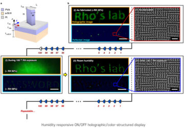
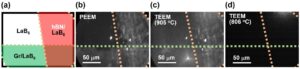
Tehnica de nanoimprimare pentru imagini holografice sensibile la umiditate
Nodul sursă: 1786485
Timestamp-ul: Decembrie 26, 2022
Primul bandaj electronic tranzitoriu accelerează vindecarea cu 30%
Nodul sursă: 1972187
Timestamp-ul: Februarie 22, 2023
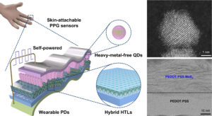
Fotosenzorul cu punct cuantic ecologic de cea mai înaltă performanță din lume, fără sursă de alimentare externă necesară
Nodul sursă: 3001715
Timestamp-ul: Decembrie 8, 2023