05 czerwca 2023 (Wiadomości Nanowerk) Chirurdzy onkolodzy będą mogli wkrótce uzyskać pełniejszy obraz nowotworów podczas operacji dzięki nowym środkom obrazowania, które mogą oświetlać wiele biomarkerów jednocześnie, podają badacze z Uniwersytetu Illinois Urbana-Champaign. Fluorescencyjne nanocząsteczki owinięte w błony czerwonych krwinek skuteczniej celują w nowotwory niż obecne klinicznie zatwierdzone barwniki i mogą emitować dwa różne sygnały w odpowiedzi na tylko jedną wiązkę światła chirurgicznego, co może pomóc lekarzom w rozróżnieniu granic guza i identyfikacji nowotworów z przerzutami .
Środki obrazujące można łączyć z kamerami inspirowanymi biologią, które naukowcy opracowali wcześniej do diagnozowania w czasie rzeczywistym podczas operacji, powiedział lider grupy badawczej Viktor Gruev, profesor inżynierii elektrycznej i komputerowej w Illinois.
W nowym badaniu opublikowanym w czasopiśmie ACS Nano ("Cell-membrane coated nanoparticles for tumor delineation and qualitative estimation of cancer biomarkers at single wavelength excitation in murine and phantom models") badacze zademonstrowali nowe nanocząstki o podwójnym sygnale w fantomach nowotworów – modelach 3D naśladujących cechy nowotworów i ich otoczenia – oraz na żywych myszach.
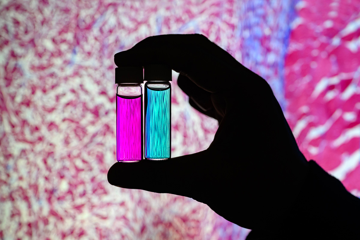 Researcher Indrajit Srivastava holds solutions of nanoparticles that can target two cancer biomarkers, giving off two distinct signals when lit by one fluorescent wavelength. This could give surgeons a more complete picture of a tumor and guide operating-room decisions. In the background is a microscopic scan of a tissue sample. (Image: Fred Zwicky)
“If you want to find all the cancer, imaging one biomarker is not enough. It could miss some tumors. If you introduce a second or a third biomarker, the likelihood of removing all cancer cells increases, and the likelihood of a better outcome for the patients increases.” said Gruev, who also is a professor in the Carle Illinois College of Medicine. “Multiple-targeted drugs and imaging agents are a recent trend, and our group is driving the trend hard because we have the camera technology that can image multiple signals at once.”
Traditionally, a surgeon removes a tumor and sends it to a pathologist for assessment, a process that can take hours to days, said Illinois postdoctoral researcher Indrajit Srivastava, the first author of the paper. As research has moved toward real-time diagnostics, several challenges have prevented wide application: Many tumor-targeted imaging agents only minimally reach their tumor targets, instead being quickly cleared from the blood stream and accumulating in the liver, Srivastava said.
“A few people before us have used nanoparticles coated with red blood cells and found they circulate longer – a few days. We saw the same thing in our mice: The membrane-coated nanoparticles circulated longer in blood, with reduced uptake in the liver. Because they were circulating longer, more of the imaging agents accumulated in the tumors, giving us a stronger fluorescent signal,” Srivastava said.
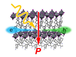
An artist’s rendering of membrane-wrapped nanoparticles binding to markers on a cancer cell and emitting colored light.
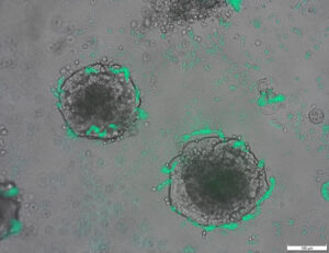
The two biomarkers targeted by the new imaging agents include one that is prevalent in early cancer and one that is prevalent in late-stage cancer, which is more likely to be metastatic. The researchers found that the probes were effective at distinguishing cancerous tissue from healthy tissue, as well as distinguishing the two signals from each other.
“This is appealing for surgical application, as it could help determine where exactly to make the cut. Having multiple signals gives a more overall picture of the tumor. And it could tell a surgeon, ‘This may be metastatic, you may want to be more aggressive in your removal.’” Srivastava said.
Only needing one wavelength of laser light to elicit multiple signals is another benefit for surgical applications, as it makes the instrumentation much more compact than those requiring multiple lasers for each needed wavelength, Gruev said.
The researchers plan to develop more tumor-imaging agents that target multiple markers, and to move forward with further preclinical and clinical studies using their dual-signal dyes with surgical goggles they have developed.
“In this battle for ensuring we remove all the cancer cells during surgery, we need investments both in the imaging camera technology and in the tumor targeting agents,” Gruev said.
Researcher Indrajit Srivastava holds solutions of nanoparticles that can target two cancer biomarkers, giving off two distinct signals when lit by one fluorescent wavelength. This could give surgeons a more complete picture of a tumor and guide operating-room decisions. In the background is a microscopic scan of a tissue sample. (Image: Fred Zwicky)
“If you want to find all the cancer, imaging one biomarker is not enough. It could miss some tumors. If you introduce a second or a third biomarker, the likelihood of removing all cancer cells increases, and the likelihood of a better outcome for the patients increases.” said Gruev, who also is a professor in the Carle Illinois College of Medicine. “Multiple-targeted drugs and imaging agents are a recent trend, and our group is driving the trend hard because we have the camera technology that can image multiple signals at once.”
Traditionally, a surgeon removes a tumor and sends it to a pathologist for assessment, a process that can take hours to days, said Illinois postdoctoral researcher Indrajit Srivastava, the first author of the paper. As research has moved toward real-time diagnostics, several challenges have prevented wide application: Many tumor-targeted imaging agents only minimally reach their tumor targets, instead being quickly cleared from the blood stream and accumulating in the liver, Srivastava said.
“A few people before us have used nanoparticles coated with red blood cells and found they circulate longer – a few days. We saw the same thing in our mice: The membrane-coated nanoparticles circulated longer in blood, with reduced uptake in the liver. Because they were circulating longer, more of the imaging agents accumulated in the tumors, giving us a stronger fluorescent signal,” Srivastava said.
An artist’s rendering of membrane-wrapped nanoparticles binding to markers on a cancer cell and emitting colored light.
The two biomarkers targeted by the new imaging agents include one that is prevalent in early cancer and one that is prevalent in late-stage cancer, which is more likely to be metastatic. The researchers found that the probes were effective at distinguishing cancerous tissue from healthy tissue, as well as distinguishing the two signals from each other.
“This is appealing for surgical application, as it could help determine where exactly to make the cut. Having multiple signals gives a more overall picture of the tumor. And it could tell a surgeon, ‘This may be metastatic, you may want to be more aggressive in your removal.’” Srivastava said.
Only needing one wavelength of laser light to elicit multiple signals is another benefit for surgical applications, as it makes the instrumentation much more compact than those requiring multiple lasers for each needed wavelength, Gruev said.
The researchers plan to develop more tumor-imaging agents that target multiple markers, and to move forward with further preclinical and clinical studies using their dual-signal dyes with surgical goggles they have developed.
“In this battle for ensuring we remove all the cancer cells during surgery, we need investments both in the imaging camera technology and in the tumor targeting agents,” Gruev said.
- Dystrybucja treści i PR oparta na SEO. Uzyskaj wzmocnienie już dziś.
- PlatoAiStream. Analiza danych Web3. Wiedza wzmocniona. Dostęp tutaj.
- Wybijanie przyszłości w Adryenn Ashley. Dostęp tutaj.
- Kupuj i sprzedawaj akcje spółek PRE-IPO z PREIPO®. Dostęp tutaj.
- Źródło: https://www.nanowerk.com/news2/biotech/newsid=63117.php
- :ma
- :Jest
- :nie
- :Gdzie
- $W GÓRĘ
- 10
- 12
- 3d
- 7
- 8
- 9
- a
- Zgromadzone
- agentów
- agresywny
- Wszystkie kategorie
- również
- an
- i
- Inne
- pociągający
- Zastosowanie
- aplikacje
- podejście
- zatwierdzony
- SĄ
- AS
- oszacowanie
- At
- autor
- tło
- Bitwa
- BE
- Belka
- bo
- zanim
- jest
- korzyści
- Ulepsz Swój
- wiążący
- biomarker
- krew
- Granice
- obie
- by
- aparat fotograficzny
- kamery
- CAN
- Rak
- Komórki nowotworowe
- Komórki
- Centrum
- wyzwania
- obiegowy
- Kliniczne
- Badania kliniczne
- bliższy
- Studentki
- połączony
- kompletny
- komputer
- Inżynieria komputerowa
- mógłby
- Aktualny
- Ciąć
- Data
- Dni
- Decyzje
- wykazać
- Ustalać
- rozwijać
- rozwinięty
- odrębny
- rozróżniać
- Lekarze
- jazdy
- Narkotyki
- podczas
- każdy
- Wcześnie
- Efektywne
- Inżynieria
- dość
- zapewnienie
- dokładnie
- Cecha
- Korzyści
- kilka
- Znajdź
- i terminów, a
- W razie zamówieenia projektu
- Naprzód
- znaleziono
- od
- dalej
- miejsce
- Dać
- daje
- Dający
- Zarządzanie
- poprowadzi
- ręka
- Ciężko
- Have
- mający
- zdrowy
- pomoc
- pomoc
- przytrzymanie
- posiada
- holistyczne
- GODZINY
- HTTPS
- zidentyfikować
- if
- Illinois
- oświetlać
- obraz
- Obrazowanie
- in
- zawierać
- Zwiększenia
- zamiast
- przedstawiać
- Inwestycje
- IT
- dziennik
- jpg
- właśnie
- tylko jeden
- laser
- Lasery
- lider
- lekki
- Prawdopodobnie
- relacja na żywo
- Wątroba
- dłużej
- robić
- WYKONUJE
- wiele
- Może..
- lekarstwo
- Myszy
- Środkowy
- modele
- jeszcze
- ruch
- pójść naprzód
- dużo
- wielokrotność
- Potrzebować
- potrzebne
- potrzeba
- Nowości
- of
- poza
- on
- pewnego razu
- ONE
- tylko
- or
- Inne
- ludzkiej,
- Wynik
- ogólny
- Papier
- pacjenci
- Ludzie
- fantom
- obraz
- krok po kroku
- plato
- Analiza danych Platona
- PlatoDane
- rozpowszechniony
- poprzednio
- wygląda tak
- Profesor
- jakościowy
- szybko
- dosięgnąć
- w czasie rzeczywistym
- zrealizować
- niedawny
- Czerwony
- Zredukowany
- usuwanie
- usunąć
- usuwanie
- wykonanie
- raport
- Badania naukowe
- Grupa poszukiwawcza
- badacz
- Badacze
- odpowiedź
- Powiedział
- taki sam
- skanować
- druga
- wysyła
- kilka
- Signal
- Sygnały
- pojedynczy
- Rozwiązania
- kilka
- Wkrótce
- strumień
- silniejszy
- badania naukowe
- Badanie
- Chirurgia
- chirurgiczny
- Brać
- biorąc
- cel
- ukierunkowane
- kierowania
- cele
- Technologia
- powiedzieć
- niż
- dzięki
- że
- Połączenia
- ich
- one
- rzecz
- Trzeci
- to
- tych
- do
- w kierunku
- tradycyjnie
- Trend
- Próby
- drugiej
- uniwersytet
- us
- używany
- za pomocą
- Zobacz i wysłuchaj
- chcieć
- we
- DOBRZE
- były
- jeśli chodzi o komunikację i motywację
- który
- KIM
- szeroki
- w
- Praca
- Owinięty
- ty
- Twój
- zefirnet
Więcej z Nanowerk
Niklowo-platynowy nanoskalowy rdzeń z platynową powłoką rozbija cząsteczki tlenu na użyteczne jony
Węzeł źródłowy: 2788122
Znak czasu: Lipiec 27, 2023
Odkrycie naprawy DNA może ulepszyć biotechnologię (w / wideo)
Węzeł źródłowy: 1987323
Znak czasu: Mar 2, 2023
Bardziej efektywny projekt eksperymentalny umożliwiający wprowadzenie komórki w nowy stan
Węzeł źródłowy: 2914286
Znak czasu: Październik 2, 2023

Naukowcy opracowują bakterie zdolne do wykrywania DNA guza
Węzeł źródłowy: 2820250
Znak czasu: Sierpnia 12, 2023

Oparta na origami integracja robotów, które wyczuwają, podejmują decyzje i reagują
Węzeł źródłowy: 2565107
Znak czasu: Kwiecień 4, 2023
Rzucenie światła na pochodzenie efektu fotowoltaicznego w perowskitach organicznych i nieorganicznych
Węzeł źródłowy: 3036090
Znak czasu: Grudnia 26, 2023
Kwantowy skok w technologii oscylatorów mechanicznych
Węzeł źródłowy: 2817575
Znak czasu: Sierpnia 11, 2023
Nowa technologia powlekania powierzchni zwiększa siedmiokrotnie emisję elektronów materiałów
Węzeł źródłowy: 2649550
Znak czasu: 12 maja 2023 r.
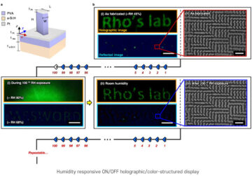
Technika nanoimprintingu dla obrazów holograficznych reagujących na wilgoć
Węzeł źródłowy: 1786485
Znak czasu: Grudnia 26, 2022
Pierwszy przejściowy bandaż elektroniczny przyspiesza gojenie o 30%
Węzeł źródłowy: 1972187
Znak czasu: Luty 22, 2023
Najbardziej wydajny na świecie, ekologiczny fotosensor z kropką kwantową, niewymagający zewnętrznego źródła zasilania
Węzeł źródłowy: 3001715
Znak czasu: Grudnia 8, 2023