05 juni 2023 (Nanowerk Nieuws) Kankerchirurgen kunnen binnenkort een vollediger beeld krijgen van tumoren tijdens operaties dankzij nieuwe beeldvormende middelen die meerdere biomarkers tegelijk kunnen verlichten, rapporteren onderzoekers van de Universiteit van Illinois, Urbana-Champaign. De fluorescerende nanodeeltjes, gewikkeld in de membranen van rode bloedcellen, richten zich beter op tumoren dan de huidige klinisch goedgekeurde kleurstoffen en kunnen twee verschillende signalen uitzenden als reactie op slechts één straal chirurgisch licht, een functie die artsen zou kunnen helpen tumorgrenzen te onderscheiden en uitgezaaide kankers te identificeren . De beeldvormende middelen kunnen worden gecombineerd met bio-geïnspireerde camera's, die de onderzoekers eerder ontwikkelden voor realtime diagnose tijdens operaties, zei onderzoeksgroepleider Viktor Gruev, een professor in elektrische en computertechniek in Illinois. In een nieuwe studie in het tijdschrift ACS Nano ("Cell-membrane coated nanoparticles for tumor delineation and qualitative estimation of cancer biomarkers at single wavelength excitation in murine and phantom models"), demonstreerden de onderzoekers hun nieuwe dual-signal nanodeeltjes in tumorfantomen - 3D-modellen die de kenmerken van tumoren en hun omgeving nabootsen - en in levende muizen.
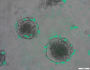
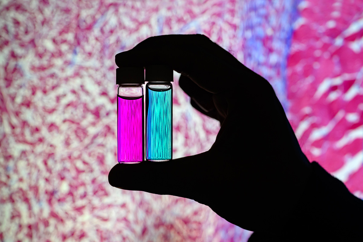 Researcher Indrajit Srivastava holds solutions of nanoparticles that can target two cancer biomarkers, giving off two distinct signals when lit by one fluorescent wavelength. This could give surgeons a more complete picture of a tumor and guide operating-room decisions. In the background is a microscopic scan of a tissue sample. (Image: Fred Zwicky)
“If you want to find all the cancer, imaging one biomarker is not enough. It could miss some tumors. If you introduce a second or a third biomarker, the likelihood of removing all cancer cells increases, and the likelihood of a better outcome for the patients increases.” said Gruev, who also is a professor in the Carle Illinois College of Medicine. “Multiple-targeted drugs and imaging agents are a recent trend, and our group is driving the trend hard because we have the camera technology that can image multiple signals at once.”
Traditionally, a surgeon removes a tumor and sends it to a pathologist for assessment, a process that can take hours to days, said Illinois postdoctoral researcher Indrajit Srivastava, the first author of the paper. As research has moved toward real-time diagnostics, several challenges have prevented wide application: Many tumor-targeted imaging agents only minimally reach their tumor targets, instead being quickly cleared from the blood stream and accumulating in the liver, Srivastava said.
“A few people before us have used nanoparticles coated with red blood cells and found they circulate longer – a few days. We saw the same thing in our mice: The membrane-coated nanoparticles circulated longer in blood, with reduced uptake in the liver. Because they were circulating longer, more of the imaging agents accumulated in the tumors, giving us a stronger fluorescent signal,” Srivastava said.
An artist’s rendering of membrane-wrapped nanoparticles binding to markers on a cancer cell and emitting colored light.
The two biomarkers targeted by the new imaging agents include one that is prevalent in early cancer and one that is prevalent in late-stage cancer, which is more likely to be metastatic. The researchers found that the probes were effective at distinguishing cancerous tissue from healthy tissue, as well as distinguishing the two signals from each other.
“This is appealing for surgical application, as it could help determine where exactly to make the cut. Having multiple signals gives a more overall picture of the tumor. And it could tell a surgeon, ‘This may be metastatic, you may want to be more aggressive in your removal.’” Srivastava said.
Only needing one wavelength of laser light to elicit multiple signals is another benefit for surgical applications, as it makes the instrumentation much more compact than those requiring multiple lasers for each needed wavelength, Gruev said.
The researchers plan to develop more tumor-imaging agents that target multiple markers, and to move forward with further preclinical and clinical studies using their dual-signal dyes with surgical goggles they have developed.
“In this battle for ensuring we remove all the cancer cells during surgery, we need investments both in the imaging camera technology and in the tumor targeting agents,” Gruev said.
Researcher Indrajit Srivastava holds solutions of nanoparticles that can target two cancer biomarkers, giving off two distinct signals when lit by one fluorescent wavelength. This could give surgeons a more complete picture of a tumor and guide operating-room decisions. In the background is a microscopic scan of a tissue sample. (Image: Fred Zwicky)
“If you want to find all the cancer, imaging one biomarker is not enough. It could miss some tumors. If you introduce a second or a third biomarker, the likelihood of removing all cancer cells increases, and the likelihood of a better outcome for the patients increases.” said Gruev, who also is a professor in the Carle Illinois College of Medicine. “Multiple-targeted drugs and imaging agents are a recent trend, and our group is driving the trend hard because we have the camera technology that can image multiple signals at once.”
Traditionally, a surgeon removes a tumor and sends it to a pathologist for assessment, a process that can take hours to days, said Illinois postdoctoral researcher Indrajit Srivastava, the first author of the paper. As research has moved toward real-time diagnostics, several challenges have prevented wide application: Many tumor-targeted imaging agents only minimally reach their tumor targets, instead being quickly cleared from the blood stream and accumulating in the liver, Srivastava said.
“A few people before us have used nanoparticles coated with red blood cells and found they circulate longer – a few days. We saw the same thing in our mice: The membrane-coated nanoparticles circulated longer in blood, with reduced uptake in the liver. Because they were circulating longer, more of the imaging agents accumulated in the tumors, giving us a stronger fluorescent signal,” Srivastava said.
An artist’s rendering of membrane-wrapped nanoparticles binding to markers on a cancer cell and emitting colored light.
The two biomarkers targeted by the new imaging agents include one that is prevalent in early cancer and one that is prevalent in late-stage cancer, which is more likely to be metastatic. The researchers found that the probes were effective at distinguishing cancerous tissue from healthy tissue, as well as distinguishing the two signals from each other.
“This is appealing for surgical application, as it could help determine where exactly to make the cut. Having multiple signals gives a more overall picture of the tumor. And it could tell a surgeon, ‘This may be metastatic, you may want to be more aggressive in your removal.’” Srivastava said.
Only needing one wavelength of laser light to elicit multiple signals is another benefit for surgical applications, as it makes the instrumentation much more compact than those requiring multiple lasers for each needed wavelength, Gruev said.
The researchers plan to develop more tumor-imaging agents that target multiple markers, and to move forward with further preclinical and clinical studies using their dual-signal dyes with surgical goggles they have developed.
“In this battle for ensuring we remove all the cancer cells during surgery, we need investments both in the imaging camera technology and in the tumor targeting agents,” Gruev said.
- Door SEO aangedreven content en PR-distributie. Word vandaag nog versterkt.
- PlatoAiStream. Web3 gegevensintelligentie. Kennis versterkt. Toegang hier.
- De toekomst slaan met Adryenn Ashley. Toegang hier.
- Koop en verkoop aandelen in PRE-IPO-bedrijven met PREIPO®. Toegang hier.
- Bron: https://www.nanowerk.com/news2/biotech/newsid=63117.php
- : heeft
- :is
- :niet
- :waar
- $UP
- 10
- 12
- 3d
- 7
- 8
- 9
- a
- Geaccumuleerd
- agenten
- agressief
- Alles
- ook
- an
- en
- Nog een
- aantrekkelijk
- Aanvraag
- toepassingen
- nadering
- goedgekeurd
- ZIJN
- AS
- beoordeling
- At
- auteur
- achtergrond
- Strijd
- BE
- Balk
- omdat
- vaardigheden
- wezen
- voordeel
- Betere
- verbindend
- biomarker
- bloed
- grenzen
- zowel
- by
- camera
- camera's
- CAN
- Kanker
- Kankercellen
- Cellen
- Centreren
- uitdagingen
- circulerende
- Klinisch
- klinische proeven
- dichterbij
- College
- gecombineerde
- compleet
- computer
- Computertechniek
- kon
- Actueel
- Snijden
- Datum
- dagen
- beslissingen
- gedemonstreerd
- Bepalen
- ontwikkelen
- ontwikkelde
- onderscheiden
- onderscheiden
- Artsen
- aandrijving
- Drugs
- gedurende
- elk
- Vroeg
- effectief
- Engineering
- genoeg
- zorgen
- precies
- Kenmerk
- Voordelen
- weinig
- VIND DE PLEK DIE PERFECT VOOR JOU IS
- Voornaam*
- Voor
- Naar voren
- gevonden
- oppompen van
- verder
- het krijgen van
- Geven
- geeft
- Vrijgevigheid
- Groep
- gids
- hand
- Hard
- Hebben
- met
- gezond
- hulp
- het helpen van
- bezit
- houdt
- holistische
- HOURS
- HTTPS
- identificeren
- if
- Illinois
- verlichten
- beeld
- Imaging
- in
- omvatten
- Verhoogt
- verkrijgen in plaats daarvan
- voorstellen
- Investeringen
- IT
- tijdschrift
- jpg
- voor slechts
- eentje maar
- laser
- lasers
- leider
- licht
- Waarschijnlijk
- leven
- Lever
- langer
- maken
- MERKEN
- veel
- Mei..
- geneeskunde
- muizen
- Midden
- modellen
- meer
- beweging
- ga vooruit
- veel
- meervoudig
- Noodzaak
- nodig
- nodig
- New
- of
- korting
- on
- eens
- EEN
- Slechts
- or
- Overige
- onze
- Resultaat
- totaal
- Papier
- patiënten
- Mensen
- spook
- beeld
- plan
- Plato
- Plato gegevensintelligentie
- PlatoData
- heersend
- die eerder
- Hoogleraar
- kwalitatieve
- snel
- bereiken
- real-time
- realiseren
- recent
- Rood
- Gereduceerd
- verwijdering
- verwijderen
- het verwijderen van
- weergave
- verslag
- onderzoek
- onderzoeksgroep
- onderzoeker
- onderzoekers
- antwoord
- Zei
- dezelfde
- aftasten
- Tweede
- verzendt
- verscheidene
- Signaal
- signalen
- single
- Oplossingen
- sommige
- Spoedig
- stream
- sterker
- studies
- Studie
- Chirurgie
- chirurgisch
- Nemen
- het nemen
- doelwit
- doelgerichte
- targeting
- doelen
- Technologie
- vertellen
- neem contact
- bedankt
- dat
- De
- hun
- ze
- ding
- Derde
- dit
- die
- naar
- in de richting van
- traditioneel
- trend
- proeven
- twee
- universiteit-
- us
- gebruikt
- gebruik
- Bekijk
- willen
- we
- GOED
- waren
- wanneer
- welke
- WIE
- breed
- Met
- Mijn werk
- Wrapped
- u
- Your
- zephyrnet
Meer van Nanowerk
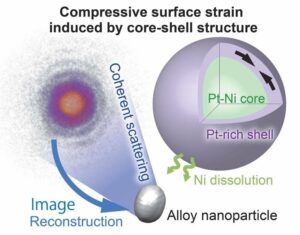
Een nikkel-platina kern op nanoschaal met een platina omhulsel kraakt zuurstofmoleculen tot bruikbare ionen
Bronknooppunt: 2788122
Tijdstempel: Juli 27, 2023
Ontdekking van DNA-reparatie kan biotechnologie verbeteren (met video)
Bronknooppunt: 1987323
Tijdstempel: 2-2023-XNUMX
Een effectiever experimenteel ontwerp om een cel in een nieuwe staat te brengen
Bronknooppunt: 2914286
Tijdstempel: Oktober 2, 2023
Onderzoekers ontwikkelen bacteriën die tumor-DNA kunnen detecteren
Bronknooppunt: 2820250
Tijdstempel: Augustus 12, 2023

Op origami gebaseerde integratie van robots die voelen, beslissen en reageren
Bronknooppunt: 2565107
Tijdstempel: 4-2023-XNUMX
Licht werpen op de oorsprong van het fotovoltaïsche effect in organisch-anorganische perovskieten
Bronknooppunt: 3036090
Tijdstempel: December 26, 2023
Een kwantumsprong in mechanische oscillatortechnologie
Bronknooppunt: 2817575
Tijdstempel: Augustus 11, 2023
Nieuwe technologie voor oppervlaktecoating verhoogt de elektronenemissie van materialen met een factor zeven
Bronknooppunt: 2649550
Tijdstempel: 12 mei 2023
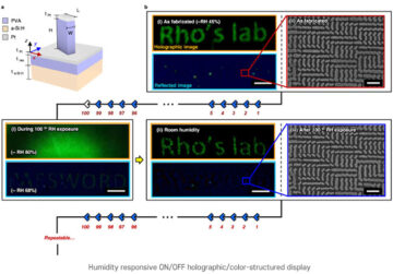
Nanoimprinting-techniek voor op vochtigheid reagerende holografische afbeeldingen
Bronknooppunt: 1786485
Tijdstempel: December 26, 2022
Eerste voorbijgaande elektronische bandage versnelt de genezing met 30%
Bronknooppunt: 1972187
Tijdstempel: Februari 22, 2023
's Werelds krachtigste, milieuvriendelijke quantum dot-fotosensor zonder dat er een externe voedingsbron nodig is
Bronknooppunt: 3001715
Tijdstempel: December 8, 2023