05 giugno 2023 (Notizie Nanowerk) I chirurghi oncologici potrebbero presto avere una visione più completa dei tumori durante l'intervento chirurgico grazie a nuovi agenti di imaging in grado di illuminare più biomarcatori contemporaneamente, riferiscono i ricercatori dell'Università dell'Illinois Urbana-Champaign. Le nanoparticelle fluorescenti, avvolte nelle membrane dei globuli rossi, prendono di mira i tumori meglio degli attuali coloranti clinicamente approvati e possono emettere due segnali distinti in risposta a un solo raggio di luce chirurgica, una caratteristica che potrebbe aiutare i medici a distinguere i confini del tumore e identificare i tumori metastatici . Gli agenti di imaging possono essere combinati con telecamere bioispirate, che i ricercatori avevano precedentemente sviluppato per la diagnosi in tempo reale durante l'intervento chirurgico, ha affermato il leader del gruppo di ricerca Viktor Gruev, professore di ingegneria elettrica e informatica dell'Illinois. In un nuovo studio sulla rivista ACS Nano ("Cell-membrane coated nanoparticles for tumor delineation and qualitative estimation of cancer biomarkers at single wavelength excitation in murine and phantom models"), i ricercatori hanno dimostrato le loro nuove nanoparticelle a doppio segnale nei fantocci tumorali - modelli 3D che imitano le caratteristiche dei tumori e dei loro dintorni - e nei topi vivi.
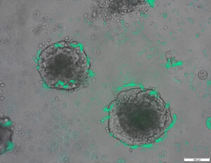
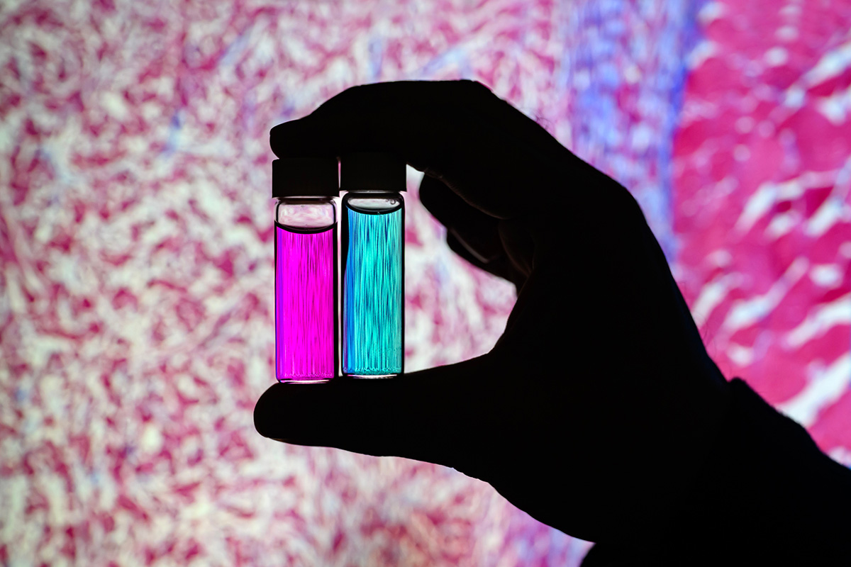 Researcher Indrajit Srivastava holds solutions of nanoparticles that can target two cancer biomarkers, giving off two distinct signals when lit by one fluorescent wavelength. This could give surgeons a more complete picture of a tumor and guide operating-room decisions. In the background is a microscopic scan of a tissue sample. (Image: Fred Zwicky)
“If you want to find all the cancer, imaging one biomarker is not enough. It could miss some tumors. If you introduce a second or a third biomarker, the likelihood of removing all cancer cells increases, and the likelihood of a better outcome for the patients increases.” said Gruev, who also is a professor in the Carle Illinois College of Medicine. “Multiple-targeted drugs and imaging agents are a recent trend, and our group is driving the trend hard because we have the camera technology that can image multiple signals at once.”
Traditionally, a surgeon removes a tumor and sends it to a pathologist for assessment, a process that can take hours to days, said Illinois postdoctoral researcher Indrajit Srivastava, the first author of the paper. As research has moved toward real-time diagnostics, several challenges have prevented wide application: Many tumor-targeted imaging agents only minimally reach their tumor targets, instead being quickly cleared from the blood stream and accumulating in the liver, Srivastava said.
“A few people before us have used nanoparticles coated with red blood cells and found they circulate longer – a few days. We saw the same thing in our mice: The membrane-coated nanoparticles circulated longer in blood, with reduced uptake in the liver. Because they were circulating longer, more of the imaging agents accumulated in the tumors, giving us a stronger fluorescent signal,” Srivastava said.
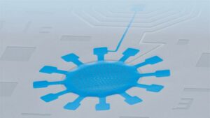
An artist’s rendering of membrane-wrapped nanoparticles binding to markers on a cancer cell and emitting colored light.
The two biomarkers targeted by the new imaging agents include one that is prevalent in early cancer and one that is prevalent in late-stage cancer, which is more likely to be metastatic. The researchers found that the probes were effective at distinguishing cancerous tissue from healthy tissue, as well as distinguishing the two signals from each other.
“This is appealing for surgical application, as it could help determine where exactly to make the cut. Having multiple signals gives a more overall picture of the tumor. And it could tell a surgeon, ‘This may be metastatic, you may want to be more aggressive in your removal.’” Srivastava said.
Only needing one wavelength of laser light to elicit multiple signals is another benefit for surgical applications, as it makes the instrumentation much more compact than those requiring multiple lasers for each needed wavelength, Gruev said.
The researchers plan to develop more tumor-imaging agents that target multiple markers, and to move forward with further preclinical and clinical studies using their dual-signal dyes with surgical goggles they have developed.
“In this battle for ensuring we remove all the cancer cells during surgery, we need investments both in the imaging camera technology and in the tumor targeting agents,” Gruev said.
Researcher Indrajit Srivastava holds solutions of nanoparticles that can target two cancer biomarkers, giving off two distinct signals when lit by one fluorescent wavelength. This could give surgeons a more complete picture of a tumor and guide operating-room decisions. In the background is a microscopic scan of a tissue sample. (Image: Fred Zwicky)
“If you want to find all the cancer, imaging one biomarker is not enough. It could miss some tumors. If you introduce a second or a third biomarker, the likelihood of removing all cancer cells increases, and the likelihood of a better outcome for the patients increases.” said Gruev, who also is a professor in the Carle Illinois College of Medicine. “Multiple-targeted drugs and imaging agents are a recent trend, and our group is driving the trend hard because we have the camera technology that can image multiple signals at once.”
Traditionally, a surgeon removes a tumor and sends it to a pathologist for assessment, a process that can take hours to days, said Illinois postdoctoral researcher Indrajit Srivastava, the first author of the paper. As research has moved toward real-time diagnostics, several challenges have prevented wide application: Many tumor-targeted imaging agents only minimally reach their tumor targets, instead being quickly cleared from the blood stream and accumulating in the liver, Srivastava said.
“A few people before us have used nanoparticles coated with red blood cells and found they circulate longer – a few days. We saw the same thing in our mice: The membrane-coated nanoparticles circulated longer in blood, with reduced uptake in the liver. Because they were circulating longer, more of the imaging agents accumulated in the tumors, giving us a stronger fluorescent signal,” Srivastava said.
An artist’s rendering of membrane-wrapped nanoparticles binding to markers on a cancer cell and emitting colored light.
The two biomarkers targeted by the new imaging agents include one that is prevalent in early cancer and one that is prevalent in late-stage cancer, which is more likely to be metastatic. The researchers found that the probes were effective at distinguishing cancerous tissue from healthy tissue, as well as distinguishing the two signals from each other.
“This is appealing for surgical application, as it could help determine where exactly to make the cut. Having multiple signals gives a more overall picture of the tumor. And it could tell a surgeon, ‘This may be metastatic, you may want to be more aggressive in your removal.’” Srivastava said.
Only needing one wavelength of laser light to elicit multiple signals is another benefit for surgical applications, as it makes the instrumentation much more compact than those requiring multiple lasers for each needed wavelength, Gruev said.
The researchers plan to develop more tumor-imaging agents that target multiple markers, and to move forward with further preclinical and clinical studies using their dual-signal dyes with surgical goggles they have developed.
“In this battle for ensuring we remove all the cancer cells during surgery, we need investments both in the imaging camera technology and in the tumor targeting agents,” Gruev said.
- Distribuzione di contenuti basati su SEO e PR. Ricevi amplificazione oggi.
- PlatoAiStream. Intelligenza dei dati Web3. Conoscenza amplificata. Accedi qui.
- Coniare il futuro con Adryenn Ashley. Accedi qui.
- Acquista e vendi azioni in società PRE-IPO con PREIPO®. Accedi qui.
- Fonte: https://www.nanowerk.com/news2/biotech/newsid=63117.php
- :ha
- :È
- :non
- :Dove
- $ SU
- 10
- 12
- 3d
- 7
- 8
- 9
- a
- Accumulato
- agenti
- aggressivo
- Tutti
- anche
- an
- ed
- Un altro
- attraente
- Applicazioni
- applicazioni
- approccio
- approvato
- SONO
- AS
- valutazione
- At
- autore
- sfondo
- Battaglia
- BE
- Larghezza
- perché
- prima
- essendo
- beneficio
- Meglio
- rilegatura
- biomarcatore
- sangue
- frontiere
- entrambi
- by
- stanza
- telecamere
- Materiale
- Cancro
- Cellule cancerogene
- Celle
- centro
- sfide
- circolante
- Info su
- test clinici
- più vicino
- College
- combinato
- completamento di una
- computer
- Ingegneria Informatica
- potuto
- Corrente
- taglio
- Data
- Giorni
- decisioni
- dimostrato
- Determinare
- sviluppare
- sviluppato
- distinto
- distinguere
- Dottori
- guida
- farmaci
- durante
- ogni
- Presto
- Efficace
- Ingegneria
- abbastanza
- assicurando
- di preciso
- caratteristica
- Caratteristiche
- pochi
- Trovare
- Nome
- Nel
- Avanti
- essere trovato
- da
- ulteriormente
- ottenere
- Dare
- dà
- Dare
- Gruppo
- guida
- cura
- Hard
- Avere
- avendo
- sano
- Aiuto
- aiutare
- possesso
- detiene
- olistica
- ORE
- HTTPS
- identificare
- if
- Illinois
- illuminare
- Immagine
- Imaging
- in
- includere
- Aumenta
- invece
- introdurre
- Investimenti
- IT
- rivista
- jpg
- ad appena
- solo uno
- laser
- Luxinar SR AOM
- leader
- leggera
- probabile
- vivere
- Fegato
- più a lungo
- make
- FA
- molti
- Maggio..
- medicina
- topi
- In mezzo
- modelli
- Scopri di più
- cambiano
- andare avanti
- molti
- multiplo
- Bisogno
- di applicazione
- che necessitano di
- New
- of
- MENO
- on
- una volta
- ONE
- esclusivamente
- or
- Altro
- nostro
- Risultato
- complessivo
- Carta
- pazienti
- Persone
- fantasma
- immagine
- piano
- Platone
- Platone Data Intelligence
- PlatoneDati
- atleta
- in precedenza
- processi
- Insegnante
- qualitativo
- rapidamente
- raggiungere
- tempo reale
- rendersi conto
- recente
- Rosso
- Ridotto
- rimozione
- rimuovere
- rimozione
- interpretazione
- rapporto
- riparazioni
- gruppo di ricerca
- ricercatore
- ricercatori
- risposta
- Suddetto
- stesso
- scansione
- Secondo
- invia
- alcuni
- Signal
- Segnali
- singolo
- Soluzioni
- alcuni
- Arrivo
- ruscello
- più forte
- studi
- Studio
- Chirurgia
- chirurgico
- Fai
- presa
- Target
- mirata
- mira
- obiettivi
- Tecnologia
- dire
- di
- Grazie
- che
- I
- loro
- di
- cosa
- Terza
- questo
- quelli
- a
- verso
- tradizionalmente
- Trend
- studi clinici
- seconda
- Università
- us
- utilizzato
- utilizzando
- Visualizza
- volere
- we
- WELL
- sono stati
- quando
- quale
- OMS
- largo
- con
- Lavora
- Avvolto
- Tu
- Trasferimento da aeroporto a Sharm
- zefiro
Di più da Nanowerk
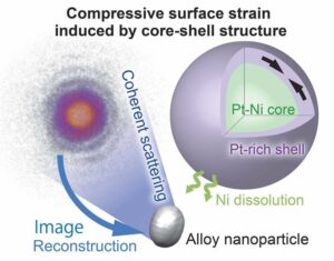
Un nucleo nanometrico di nichel-platino con un guscio di platino scompone le molecole di ossigeno in ioni utili
Nodo di origine: 2788122
Timestamp: Luglio 27, 2023
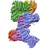
La scoperta della riparazione del DNA potrebbe migliorare la biotecnologia (con video)
Nodo di origine: 1987323
Timestamp: marzo 2, 2023
Un progetto sperimentale più efficace per ingegnerizzare una cellula in un nuovo stato
Nodo di origine: 2914286
Timestamp: Ottobre 2, 2023

I ricercatori progettano batteri in grado di rilevare il DNA tumorale
Nodo di origine: 2820250
Timestamp: 12 agosto 2023

Integrazione basata su origami di robot che percepiscono, decidono e rispondono
Nodo di origine: 2565107
Timestamp: APRILE 4, 2023
Far luce sull'origine dell'effetto fotovoltaico nelle perovskiti organico-inorganiche
Nodo di origine: 3036090
Timestamp: Dicembre 26, 2023
Un salto di qualità nella tecnologia degli oscillatori meccanici
Nodo di origine: 2817575
Timestamp: 11 agosto 2023
La nuova tecnologia di rivestimento superficiale aumenta di sette volte l'emissione di elettroni dei materiali
Nodo di origine: 2649550
Timestamp: 12 Maggio 2023
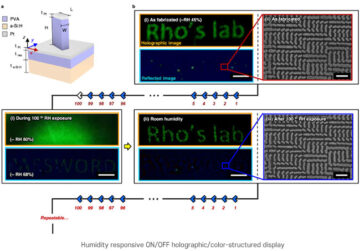
Tecnica di nanoimprinting per immagini olografiche sensibili all'umidità
Nodo di origine: 1786485
Timestamp: Dicembre 26, 2022
Il primo bendaggio elettronico transitorio accelera la guarigione del 30%
Nodo di origine: 1972187
Timestamp: Febbraio 22, 2023
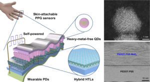
Il fotosensore a punti quantici ecologico più performante al mondo che non richiede alcuna fonte di alimentazione esterna
Nodo di origine: 3001715
Timestamp: Dicembre 8, 2023