05 juin 2023 (Actualités Nanowerk) Les chirurgiens du cancer pourraient bientôt avoir une vue plus complète des tumeurs pendant la chirurgie grâce à de nouveaux agents d'imagerie capables d'éclairer plusieurs biomarqueurs à la fois, rapportent des chercheurs de l'Université de l'Illinois à Urbana-Champaign. Les nanoparticules fluorescentes, enveloppées dans les membranes des globules rouges, ciblent mieux les tumeurs que les colorants actuellement approuvés cliniquement et peuvent émettre deux signaux distincts en réponse à un seul faisceau de lumière chirurgicale, une caractéristique qui pourrait aider les médecins à distinguer les frontières tumorales et à identifier les cancers métastatiques . Les agents d'imagerie peuvent être combinés avec des caméras bio-inspirées, que les chercheurs ont précédemment développées pour le diagnostic en temps réel pendant la chirurgie, a déclaré le chef du groupe de recherche Viktor Gruev, professeur de génie électrique et informatique dans l'Illinois. Dans une nouvelle étude publiée dans la revue ACS Nano ("Cell-membrane coated nanoparticles for tumor delineation and qualitative estimation of cancer biomarkers at single wavelength excitation in murine and phantom models"), les chercheurs ont démontré leurs nouvelles nanoparticules à double signal dans des fantômes tumoraux – des modèles 3D qui imitent les caractéristiques des tumeurs et de leur environnement – et chez des souris vivantes.
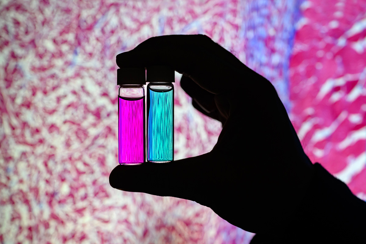 Researcher Indrajit Srivastava holds solutions of nanoparticles that can target two cancer biomarkers, giving off two distinct signals when lit by one fluorescent wavelength. This could give surgeons a more complete picture of a tumor and guide operating-room decisions. In the background is a microscopic scan of a tissue sample. (Image: Fred Zwicky)
“If you want to find all the cancer, imaging one biomarker is not enough. It could miss some tumors. If you introduce a second or a third biomarker, the likelihood of removing all cancer cells increases, and the likelihood of a better outcome for the patients increases.” said Gruev, who also is a professor in the Carle Illinois College of Medicine. “Multiple-targeted drugs and imaging agents are a recent trend, and our group is driving the trend hard because we have the camera technology that can image multiple signals at once.”
Traditionally, a surgeon removes a tumor and sends it to a pathologist for assessment, a process that can take hours to days, said Illinois postdoctoral researcher Indrajit Srivastava, the first author of the paper. As research has moved toward real-time diagnostics, several challenges have prevented wide application: Many tumor-targeted imaging agents only minimally reach their tumor targets, instead being quickly cleared from the blood stream and accumulating in the liver, Srivastava said.
“A few people before us have used nanoparticles coated with red blood cells and found they circulate longer – a few days. We saw the same thing in our mice: The membrane-coated nanoparticles circulated longer in blood, with reduced uptake in the liver. Because they were circulating longer, more of the imaging agents accumulated in the tumors, giving us a stronger fluorescent signal,” Srivastava said.
An artist’s rendering of membrane-wrapped nanoparticles binding to markers on a cancer cell and emitting colored light.
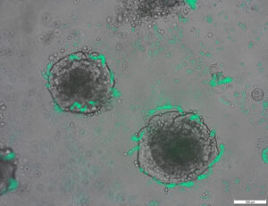
The two biomarkers targeted by the new imaging agents include one that is prevalent in early cancer and one that is prevalent in late-stage cancer, which is more likely to be metastatic. The researchers found that the probes were effective at distinguishing cancerous tissue from healthy tissue, as well as distinguishing the two signals from each other.
“This is appealing for surgical application, as it could help determine where exactly to make the cut. Having multiple signals gives a more overall picture of the tumor. And it could tell a surgeon, ‘This may be metastatic, you may want to be more aggressive in your removal.’” Srivastava said.
Only needing one wavelength of laser light to elicit multiple signals is another benefit for surgical applications, as it makes the instrumentation much more compact than those requiring multiple lasers for each needed wavelength, Gruev said.
The researchers plan to develop more tumor-imaging agents that target multiple markers, and to move forward with further preclinical and clinical studies using their dual-signal dyes with surgical goggles they have developed.
“In this battle for ensuring we remove all the cancer cells during surgery, we need investments both in the imaging camera technology and in the tumor targeting agents,” Gruev said.
Researcher Indrajit Srivastava holds solutions of nanoparticles that can target two cancer biomarkers, giving off two distinct signals when lit by one fluorescent wavelength. This could give surgeons a more complete picture of a tumor and guide operating-room decisions. In the background is a microscopic scan of a tissue sample. (Image: Fred Zwicky)
“If you want to find all the cancer, imaging one biomarker is not enough. It could miss some tumors. If you introduce a second or a third biomarker, the likelihood of removing all cancer cells increases, and the likelihood of a better outcome for the patients increases.” said Gruev, who also is a professor in the Carle Illinois College of Medicine. “Multiple-targeted drugs and imaging agents are a recent trend, and our group is driving the trend hard because we have the camera technology that can image multiple signals at once.”
Traditionally, a surgeon removes a tumor and sends it to a pathologist for assessment, a process that can take hours to days, said Illinois postdoctoral researcher Indrajit Srivastava, the first author of the paper. As research has moved toward real-time diagnostics, several challenges have prevented wide application: Many tumor-targeted imaging agents only minimally reach their tumor targets, instead being quickly cleared from the blood stream and accumulating in the liver, Srivastava said.
“A few people before us have used nanoparticles coated with red blood cells and found they circulate longer – a few days. We saw the same thing in our mice: The membrane-coated nanoparticles circulated longer in blood, with reduced uptake in the liver. Because they were circulating longer, more of the imaging agents accumulated in the tumors, giving us a stronger fluorescent signal,” Srivastava said.
An artist’s rendering of membrane-wrapped nanoparticles binding to markers on a cancer cell and emitting colored light.
The two biomarkers targeted by the new imaging agents include one that is prevalent in early cancer and one that is prevalent in late-stage cancer, which is more likely to be metastatic. The researchers found that the probes were effective at distinguishing cancerous tissue from healthy tissue, as well as distinguishing the two signals from each other.
“This is appealing for surgical application, as it could help determine where exactly to make the cut. Having multiple signals gives a more overall picture of the tumor. And it could tell a surgeon, ‘This may be metastatic, you may want to be more aggressive in your removal.’” Srivastava said.
Only needing one wavelength of laser light to elicit multiple signals is another benefit for surgical applications, as it makes the instrumentation much more compact than those requiring multiple lasers for each needed wavelength, Gruev said.
The researchers plan to develop more tumor-imaging agents that target multiple markers, and to move forward with further preclinical and clinical studies using their dual-signal dyes with surgical goggles they have developed.
“In this battle for ensuring we remove all the cancer cells during surgery, we need investments both in the imaging camera technology and in the tumor targeting agents,” Gruev said.
- Contenu propulsé par le référencement et distribution de relations publiques. Soyez amplifié aujourd'hui.
- PlatoAiStream. Intelligence des données Web3. Connaissance Amplifiée. Accéder ici.
- Frapper l'avenir avec Adryenn Ashley. Accéder ici.
- Achetez et vendez des actions de sociétés PRE-IPO avec PREIPO®. Accéder ici.
- La source: https://www.nanowerk.com/news2/biotech/newsid=63117.php
- :possède
- :est
- :ne pas
- :où
- $UP
- 10
- 12
- 3d
- 7
- 8
- 9
- a
- Accumulé
- agents
- agressif
- Tous
- aussi
- an
- ainsi que
- Une autre
- attirant
- Candidature
- applications
- une approche
- ,
- SONT
- AS
- Évaluation de risque climatique
- At
- auteur
- fond
- Bataille
- BE
- Faisceau
- car
- before
- va
- profiter
- Améliorée
- propriétés de liant
- biomarqueur
- sang
- limites
- tous les deux
- by
- appareil photo
- de CAMÉRAS de surveillance
- CAN
- Cancer
- Cellules cancéreuses
- Cellules
- Canaux centraux
- globaux
- circulé
- Infos sur les
- essais cliniques
- plus
- Université
- combiné
- complet
- ordinateur
- Ingénierie informatique
- pourriez
- Courant
- Cut/Taille
- Date
- jours
- décisions
- démontré
- Déterminer
- développer
- développé
- distinct
- distinguer
- Docteur
- conduite
- Médicaments
- pendant
- chacun
- "Early Bird"
- Efficace
- ENGINEERING
- assez
- assurer
- exactement
- Fonctionnalité
- Fonctionnalités:
- few
- Trouvez
- Prénom
- Pour
- Avant
- trouvé
- de
- plus
- obtention
- Donner
- donne
- Don
- Réservation de groupe
- guide
- main
- Dur
- Vous avez
- ayant
- la santé
- aider
- aider
- tenue
- détient
- holistique
- HEURES
- HTTPS
- identifier
- if
- Illinois
- éclairer
- image
- Imagerie
- in
- comprendre
- Augmente
- plutôt ;
- introduire
- Investissements
- IT
- Journal
- jpg
- juste
- juste un
- laser
- Luxinar SR AOM
- leader
- lumière
- Probable
- le travail
- Foie
- plus long
- faire
- FAIT DU
- de nombreuses
- Mai..
- médecine
- souris
- Milieu
- numériques jumeaux (digital twin models)
- PLUS
- Bougez
- Avance
- beaucoup
- plusieurs
- Besoin
- nécessaire
- besoin
- Nouveauté
- of
- de rabais
- on
- une fois
- ONE
- uniquement
- or
- Autre
- nos
- Résultat
- global
- Papier
- patients
- Personnes
- fantôme
- image
- plan
- Platon
- Intelligence des données Platon
- PlatonDonnées
- répandue
- précédemment
- processus
- Professeur
- qualitatif
- vite.
- nous joindre
- en temps réel
- réaliser
- récent
- Rouge
- Prix Réduit
- enlèvement
- supprimez
- enlever
- rendu
- rapport
- un article
- groupe de recherche
- chercheur
- chercheurs
- réponse
- Saïd
- même
- balayage
- Deuxièmement
- envoie
- plusieurs
- Signal
- signaux
- unique
- Solutions
- quelques
- disponible
- courant
- plus efficacement
- et le cannabis
- Étude
- #
- chirurgical
- Prenez
- prise
- Target
- des campagnes marketing ciblées,
- ciblage
- objectifs
- Technologie
- dire
- que
- à
- qui
- La
- leur
- l'ont
- chose
- Troisièmement
- this
- ceux
- à
- vers
- traditionnellement
- Trend
- essais cliniques
- deux
- université
- us
- d'utiliser
- en utilisant
- Voir
- souhaitez
- we
- WELL
- ont été
- quand
- qui
- WHO
- large
- avec
- Activités:
- Enveloppé
- you
- Votre
- zéphyrnet
Plus de Nanowerk
Un noyau nanométrique en nickel-platine avec une coque en platine craque les molécules d'oxygène en ions utiles
Nœud source: 2788122
Horodatage: Le 27 juillet 2023
La découverte de la réparation de l'ADN pourrait améliorer la biotechnologie (avec vidéo)
Nœud source: 1987323
Horodatage: 2 Mar 2023
Une conception expérimentale plus efficace pour transformer une cellule en un nouvel état
Nœud source: 2914286
Horodatage: Le 2 octobre 2023

Des chercheurs créent des bactéries capables de détecter l'ADN tumoral
Nœud source: 2820250
Horodatage: 12 août 2023

Intégration basée sur l'origami de robots qui détectent, décident et réagissent
Nœud source: 2565107
Horodatage: 4 avril 2023
Faire la lumière sur l’origine de l’effet photovoltaïque dans les pérovskites organiques-inorganiques
Nœud source: 3036090
Horodatage: Le 26 décembre 2023
Un saut quantique dans la technologie des oscillateurs mécaniques
Nœud source: 2817575
Horodatage: 11 août 2023
La nouvelle technologie de revêtement de surface multiplie par sept l'émission d'électrons des matériaux
Nœud source: 2649550
Horodatage: 12 mai 2023
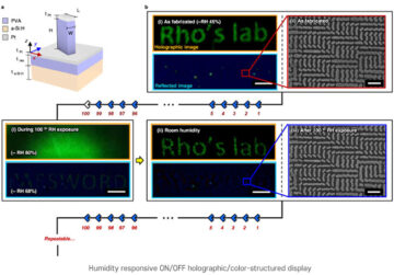
Technique de nanoimpression pour des images holographiques sensibles à l'humidité
Nœud source: 1786485
Horodatage: Le 26 décembre 2022
Le premier bandage électronique transitoire accélère la guérison de 30 %
Nœud source: 1972187
Horodatage: 22 février 2023
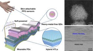
Le photocapteur à points quantiques écologique le plus performant au monde, sans source d'alimentation externe requise
Nœud source: 3001715
Horodatage: Le 8 décembre 2023